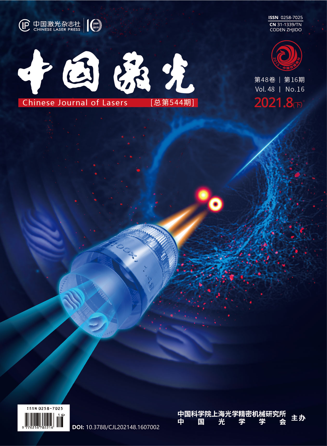
共路并行荧光辐射差分超分辨显微成像  下载: 952次封面文章
Objective Fluorescence emission difference (FED) microscopy is a versatile super-resolution microscopy flexible for all types of fluorescent dyes. However, the limited imaging speed is the main drawback that hinders the application of FED, owing to the double imaging process of positive confocal image using a solid spot scan and negative confocal image using a negative spot scan. Parallel fluorescence microscopy can overcome this limitation owing to its ability to simultaneously capture positive and negative confocal images. However, the complexity of this method’s system will increase instability and difficulties in daily maintenance, which also considerably restricts the popularization of the method. In this study, a new imaging method using a spatial light modulator (SLM) named common-path parallel FED (cpFED) microscopy was proposed. Compared with the traditional parallel FED microscopy, the proposed method that uses SLM and common path modulation maintains the advantage of doubling the imaging speed while overcoming the impact of instabilities introduced by different devices in noncommon-path parallel systems and simplifies the light path.
Methods A property of SLM is that only linear polarized light can be modulated in a fixed direction, which is the major property of common-path parallel FED microscopy. Using the polarization rotation realized by passing forth and back through a quarter-wave plate, SLM can modulate the s and p polarization components of the emitted light (Fig. 1). Using two-phase grayscale patterns, vortex and tilt-grating modulation patterns, to simultaneously modulate the horizontal and vertical polarization components of the incident laser beam on a single SLM, a staggering Gaussian solid spot and a hollow spot are generated to form the final convergent light field at the focus plane. The solid and hollow spots scan the sample simultaneously, and the fluorescence signal excited by the two spots is measured using two detectors, which will introduce a fixed transverse displacement between two images. Translating the positive confocal image to align with the negative confocal image and combining with the FED algorithm, the fast super-resolution imaging of the sample is realized. Experimental results show that the proposed method exhibits good ultradiffraction-limit imaging capabilities and high imaging speed and the resolution can be increased by approximately two times compared with traditional confocal imaging.
Results and Discussions To test the performance of cpFED, the experimental results of 200 nm and 100 nm fluorescent beads were presented herein. The 200 nm fluorescent beads were used to adjust the system and demonstrate the translation effect between the positive and negative confocal images because of its’ high fluorescent quantum efficiency (Fig. 2). The 100 nm fluorescent beads were used to measure and determine the optimum resolution performance of cpFED (Fig. 3). The full width at half maximum of a single bead plotted in Fig. 3(c) reveals that the resolution of cpFED in our system can reach up to 133 nm, indicating that the resolution of cpFED can be increased by approximately two times compared with traditional confocal imaging. Furthermore, a vimentin sample was used to verify the biological application of cpFED. The insects depicted in Fig. 4(a1), (b1), (a2), and (b2) show that the resolution and contrast in cpFED can be significantly promoted, while the noise in cpFED is considerably lower than that in traditional confocal imaging.
Conclusions Using SLM, cpFED can realize a small translation between solid and donut spots in a common path and simultaneously capture positive and negative confocal images, thus overcoming the imaging speed limit and promoting the biological application of FED. Compared with pFED, the proposed cpFED simplifies the system structure, reduces the adjustment complexity, and improves the robustness of the system because of the SLM flexibility. Experimental results demonstrate that cpFED achieves a resolution improvement of two times that of traditional confocal imaging while achieving double the imaging speed.
张智敏, 黄宇然, 刘少聪, 匡翠方, 曹良才, 刘勇, 韩于冰, 郝翔, 刘旭. 共路并行荧光辐射差分超分辨显微成像[J]. 中国激光, 2021, 48(16): 1607002. Zhimin Zhang, Yuran Huang, Shaocong Liu, Cuifang Kuang, Liangcai Cao, Yong Liu, Yubing Han, Xiang Hao, Xu Liu. Common-Path Parallel Fluorescence Emission Difference Super-Resolution Microscopy[J]. Chinese Journal of Lasers, 2021, 48(16): 1607002.


