Photonics Research, 2020, 8 (12): 12001875, Published Online: Nov. 19, 2020  

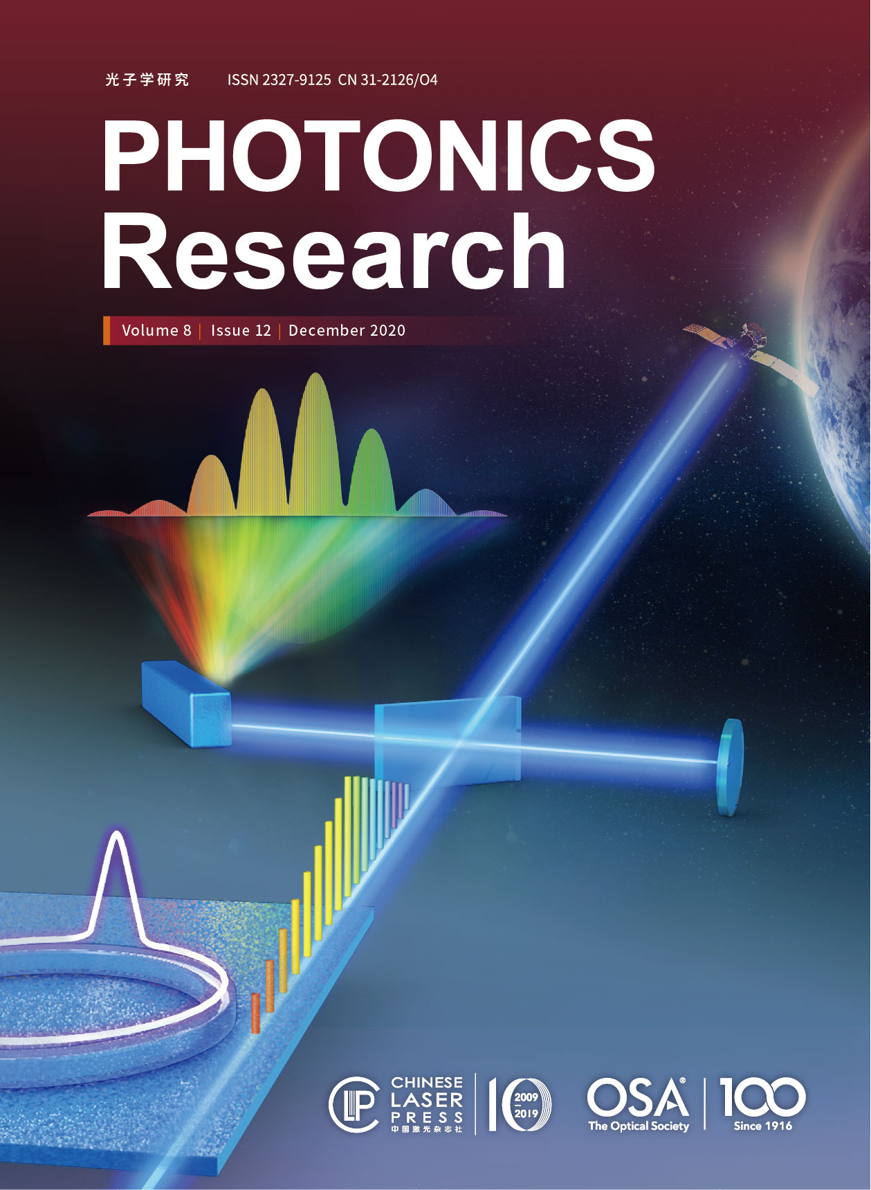
Confocal visible/NIR photoacoustic microscopy of tumors with structural, functional, and nanoprobe contrasts Download: 720次

Author Affiliations
1 Department of Biomedical Engineering, City University of Hong Kong, 999077 Hong Kong SAR, China
2 Center of Super-Diamond and Advanced Films (COSDAF) and Department of Chemistry, City University of Hong Kong, 999077 Hong Kong SAR, China
3 City University of Hong Kong Shenzhen Research Institute, Shenzhen 518057, China
4 e-mail: apcslee@cityu.edu.hk
5 e-mail: lidawang@cityu.edu.hk
Copy Citation Text

Jiangbo Chen, Yachao Zhang, Xiaozhen Li, Jingyi Zhu, Dengfeng Li, Shengliang Li, Chun-Sing Lee, Lidai Wang. Confocal visible/NIR photoacoustic microscopy of tumors with structural, functional, and nanoprobe contrasts[J]. Photonics Research, 2020, 8(12): 12001875.

References

[1] F. A. de la Peña, R. Andrés, J. A. Garcia-Sáenz, L. Manso, M. Margelí, E. Dalmau, S. Pernas, A. Prat, S. Servitja, E. Ciruelos. SEOM clinical guidelines in early stage breast cancer (2018). Clin. Transl. Oncol., 2019, 21: 18-30.

[2] F. Cardoso, S. Kyriakides, S. Ohno, F. Penault-Llorca, P. Poortmans, I. T. Rubio, S. Zackrisson, E. Senkus. Early breast cancer: ESMO clinical practice guidelines for diagnosis, treatment and follow-up. Ann. Oncol., 2019, 30: 1194-1220.

[3] K. Anand, J. Roszik, D. Gombos, J. Upshaw, V. Sarli, S. Meas, A. Lucci, C. Hall, S. Patel. Pilot study of circulating tumor cells in early-stage and metastatic uveal melanoma. Cancers, 2019, 11: 856.

[4] S. Cheraghlou, S. R. Christensen, G. O. Agogo, M. Girardi. Comparison of survival after mohs micrographic surgery vs wide margin excision for early-stage invasive melanoma. JAMA Dermatol., 2019, 155: 1252-1259.

[5] C. Sompong, S. Wongthanavasu. An efficient brain tumor segmentation based on cellular automata and improved tumor-cut algorithm. Exp. Syst. Appl., 2017, 72: 231-244.

[6] C. Yeh, L. Wang, J. Liang, Y. Zhou, S. Hu, R. E. Sohn, J. M. Arbeit, L. V. Wang. Early-stage tumor detection using photoacoustic microscopy: a pattern recognition approach. Proc. SPIE, 2017, 10064: 100644N.

[7] A. Binte, E. Attia, G. Balasundaram, M. Moothanchery, U. S. Dinish, R. Bi, V. Ntziachristos, M. Olivo. A review of clinical photoacoustic imaging: current and future trends. Photoacoustics, 2019, 16: 100144.

[8] S. Manohar, S. S. Gambhir. Clinical photoacoustic imaging. Photoacoustics, 2020, 19: 100196.

[9] H. C. Zhou, N. Chen, H. Zhao, T. Yin, J. Zhang, W. Zheng, L. Song, C. Liu, R. Zheng. Optical-resolution photoacoustic microscopy for monitoring vascular normalization during anti-angiogenic therapy. Photoacoustics, 2019, 15: 100143.

[10] W. Zhang, Y. Li, Y. Yu, K. Derouin, Y. Qin, V. P. Nguyen, X. Xia, X. Wang, Y. M. Paulus. Simultaneous photoacoustic microscopy, spectral-domain optical coherence tomography, and fluorescein microscopy multi-modality retinal imaging. Photoacoustics, 2020, 20: 100194.

[11] Y. Duan, D. Hu, B. Guo, Q. Shi, M. Wu, S. Xu, X. Liu, J. Jiang, Z. Sheng, H. Zheng, B. Liu. Nanostructural control enables optimized photoacoustic-fluorescence–magnetic resonance multimodal imaging and photothermal therapy of brain tumor. Adv. Funct. Mater., 2020, 30: 1907077.

[12] B. Guo, Z. Feng, D. Hu, S. Xu, E. Middha, Y. Pan, C. Liu, H. Zheng, J. Qian, Z. Sheng, B. Liu. Precise deciphering of brain vasculatures and microscopic tumors with dual NIR-II fluorescence and photoacoustic imaging. Adv. Mater., 2019, 31: 1902504.

[13] G. S. Filonov, A. Krumholz, J. Xia, J. Yao, L. V. Wang, V. V. Verkhusha. Deep-tissue photoacoustic tomography of a genetically encoded near-infrared fluorescent probe. Angew. Chem. Int. Ed., 2012, 51: 1448-1451.

[14] C. Liu, D. Wang, Y. Zhan, L. Yan, Q. Lu, M. Y. Z. Chang, J. Luo, L. Wang, D. Du, Y. Lin, J. Xia, Y. Wu. Switchable photoacoustic imaging of glutathione using MnO2 nanotubes for cancer diagnosis. ACS Appl. Mater. Interfaces, 2018, 10: 44231-44239.

[15] N. Nyayapathi, J. Xia. Photoacoustic imaging of breast cancer: a mini review of system design and image features. J. Biomed. Opt., 2019, 24: 121911.

[16] P. K. Upputuri, M. Pramanik. Recent advances in photoacoustic contrast agents for in vivo imaging. Wiley Interdiscip. Rev. Nanomed. Nanobiotechnol., 2020, 12: e1618.

[17] W. Zhang, Y. Wen, D. X. He, Y. F. Wang, X. L. Liu, C. Li, X. J. Liang. Near-infrared AIEgens as transformers to enhance tumor treatment efficacy with controllable self-assembled redox-responsive carrier-free nanodrug. Biomaterials, 2019, 193: 12-21.

[18] G. Wen, X. Li, Y. Zhang, X. Han, X. Xu, C. Liu, K. Chan, C.-S. Lee, C. Yin, L. Bian, L. Wang. Effective phototheranostics of brain tumor assisted by near-infrared-II light-responsive semiconducting polymer nanoparticles. ACS Appl. Mater. Interfaces, 2020, 12: 33492-33499.

[19] A. Ron, X. L. Deán-Ben, S. Gottschalk, D. Razansky. Volumetric optoacoustic imaging unveils high-resolution patterns of acute and cyclic hypoxia in a murine model of breast cancer. Cancer Res., 2019, 79: 4767-4775.

[20] J. Yao. Wide-field fast-scanning photoacoustic microscopy based on a water-immersible MEMS scanning mirror. J. Biomed. Opt., 2012, 17: 080505.

[21] L. Wang, K. Maslov, L. V. Wang. Single-cell label-free photoacoustic flowoxigraphy in vivo. Proc. Natl. Acad. Sci. USA, 2013, 110: 5759-5764.

[22] L. Wang, K. Maslov, J. Yao, B. Rao, L. V. Wang. Fast voice-coil scanning optical-resolution photoacoustic microscopy. Opt. Lett., 2011, 36: 139-141.

[23] J. Kim, J. Y. Kim, S. Jeon, J. W. Baik, S. H. Cho, C. Kim. Super-resolution localization photoacoustic microscopy using intrinsic red blood cells as contrast absorbers. Light Sci. Appl., 2019, 8: 103.

[24] J. Shi, T. T. W. Wong, Y. He, L. Li, R. Zhang, C. S. Yung, J. Hwang, K. Maslov, L. V. Wang. High-resolution, high-contrast mid-infrared imaging of fresh biological samples with ultraviolet-localized photoacoustic microscopy. Nat. Photonics, 2019, 13: 609-615.

[25] J. Chen, Y. Zhang, L. He, Y. Liang, L. Wang. Wide-field polygon-scanning photoacoustic microscopy of oxygen saturation at 1-MHz A-line rate. Photoacoustics, 2020, 20: 100195.

[26] R. Cao, J. Li, B. Ning, N. Sun, T. Wang, Z. Zuo, S. Hu. Functional and oxygen-metabolic photoacoustic microscopy of the awake mouse brain. Neuroimage, 2017, 150: 77-87.

[27] D. Li, C. Liu, Y. Yang, L. Wang, Y. Shen. Micro-rocket robot with all-optic actuating and tracking in the blood. Light Sci. Appl., 2020, 9: 84.

[28] L. Wang, C. Zhang, L. V. Wang. Grueneisen relaxation photoacoustic microscopy. Phys. Rev. Lett., 2014, 113: 174301.

[29] S. Li, Q. Deng, Y. Zhang, X. Li, G. Wen, X. Cui, Y. Wan, Y. Huang, J. Chen, Z. Liu, L. Wang, C. S. Lee. Rational design of conjugated small molecules for superior photothermal theranostics in the NIR-II biowindow. Adv. Mater., 2020, 32: 2001146.

[30] M. Li, Y. Tang, J. Yao. Photoacoustic tomography of blood oxygenation: a mini review. Photoacoustics, 2018, 10: 65-73.

[31] Y. Liang, L. Jin, B.-O. Guan, L. Wang. 2  MHz multi-wavelength pulsed laser for functional photoacoustic microscopy. Opt. Lett., 2017, 42: 1452-1455.

[32] C. Liu, Y. Liang, L. Wang. Single-shot photoacoustic microscopy of hemoglobin concentration, oxygen saturation, and blood flow in sub-microseconds. Photoacoustics, 2020, 17: 100156.

[33] J. Yao, K. I. Maslov, Y. Zhang, Y. Xia, L. V. Wang. Label-free oxygen-metabolic photoacoustic microscopy in vivo. J. Biomed. Opt., 2011, 16: 076003.

[34] B. Guo, J. Chen, N. Chen, E. Middha, S. Xu, Y. Pan, M. Wu. High-resolution 3D NIR-II photoacoustic imaging of cerebral and tumor vasculatures using conjugated polymer nanoparticles as contrast agent. Adv. Mater., 2019, 31: 1808355.

[35] R. García-Álvarez, L. Chen, A. Nedilko, A. Sánchez-Iglesias, A. Rix, W. Lederle, V. Pathak, T. Lammers, G. Von Plessen, K. Kostarelos, L. M. Liz-Marzán, A. J. C. Kuehne, D. N. Chigrin. Optimizing the geometry of photoacoustically active gold nanoparticles for biomedical imaging. ACS Photon., 2020, 7: 646-652.

[36] C. Li, J. Shi, X. Wang, B. Wang, X. Gong, L. Song, K. K. Y. Wong. High-energy all-fiber gain-switched thulium-doped fiber laser for volumetric photoacoustic imaging of lipids. Photon. Res., 2020, 8: 160-164.

[37] S. Jeon, J. Kim, D. Lee, J. W. Baik, C. Kim. Review on practical photoacoustic microscopy. Photoacoustics, 2019, 15: 100141.

[38] Y. Wang, K. Maslov, C. Kim, S. Hu, L. V. Wang. Integrated photoacoustic and fluorescence confocal microscopy. IEEE Trans. Biomed. Eng., 2010, 57: 2576-2578.

[39] J. Yao, L. V. Wang. Photoacoustic microscopy. Laser Photon. Rev., 2013, 7: 758-778.

[40] L. Lin, J. Yao, R. Zhang, C. C. Chen, C. H. Huang, Y. Li, L. Wang, W. Chapman, J. Zou, L. V. Wang. High-speed photoacoustic microscopy of mouse cortical microhemodynamics. J. Biophoton., 2017, 10: 792-798.

[41] C. Stapf, J. P. Mohr, J. Pile-Spellman, R. R. Sciacca, A. Hartmann, H. C. Schumacher, H. Mast. Concurrent arterial aneurysms in brain arteriovenous malformations with haemorrhagic presentation. J. Neurol. Neurosurg. Psychiatry, 2002, 73: 294-298.

[42] A. K. Iyer, G. Khaled, J. Fang, H. Maeda. Exploiting the enhanced permeability and retention effect for tumor targeting. Drug Discov. Today, 2006, 11: 812-818.

[43] Y. Matsumura, H. Maeda. A new concept for macromolecular therapeutics in cancer chemotherapy: mechanism of tumoritropic accumulation of proteins and the antitumor agent smancs. Cancer Res., 1986, 46: 6387-6392.

Jiangbo Chen, Yachao Zhang, Xiaozhen Li, Jingyi Zhu, Dengfeng Li, Shengliang Li, Chun-Sing Lee, Lidai Wang. Confocal visible/NIR photoacoustic microscopy of tumors with structural, functional, and nanoprobe contrasts[J]. Photonics Research, 2020, 8(12): 12001875.
引用该论文: TXT   |   EndNote

相关论文

加载中...

关于本站 Cookie 的使用提示

中国光学期刊网使用基于 cookie 的技术来更好地为您提供各项服务,点击此处了解我们的隐私策略。 如您需继续使用本网站,请您授权我们使用本地 cookie 来保存部分信息。
全站搜索
您最值得信赖的光电行业旗舰网络服务平台!