光学学报, 2019, 39 (2): 0211004, 网络出版: 2019-05-10   

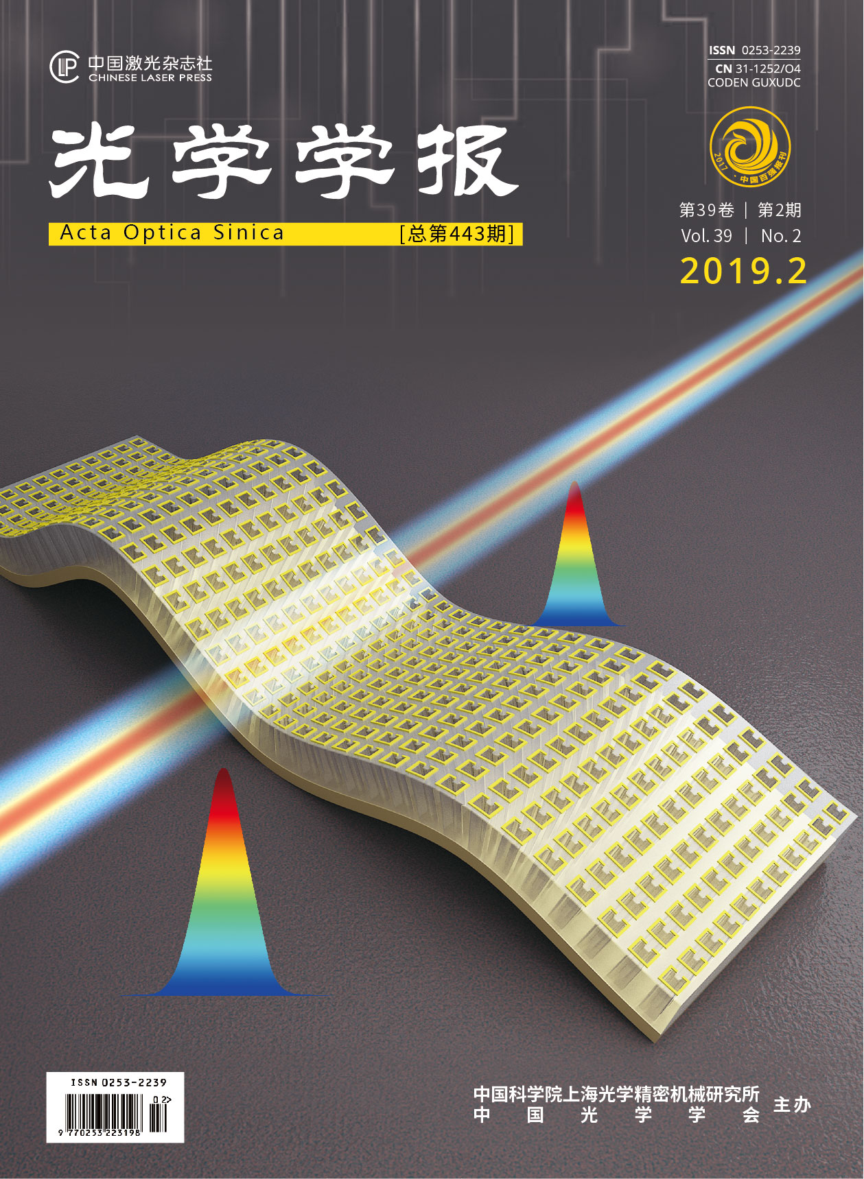
谱域相位显微成像的相位解包裹 下载: 1028次

Phase Unwrapping in Spectral Domain Phase Microscopy
作者单位
1 东北大学秦皇岛分校实验教育中心, 河北 秦皇岛 066004
2 燕山大学电气工程学院, 河北 秦皇岛 066004
3 东北大学秦皇岛分校控制工程学院, 河北 秦皇岛 066004
引用该论文

周红仙, 朱礼达, 赵玉倩, 马振鹤, 王毅. 谱域相位显微成像的相位解包裹[J]. 光学学报, 2019, 39(2): 0211004.

Hongxian Zhou, Lida Zhu, Yuqian Zhao, Zhenhe Ma, Yi Wang. Phase Unwrapping in Spectral Domain Phase Microscopy[J]. Acta Optica Sinica, 2019, 39(2): 0211004.

参考文献

[1] 王毅, 郭哲, 朱立达, 等. 基于谱域相位分辨光学相干层析的纳米级表面形貌成像[J]. 物理学报, 2017, 66(15): 154202.

    Wang Y, Guo Z, Zhu L D, et al. Nanoscale surface topography imaging using phase-resolved spectral domain optical coherence tomography[J]. Acta Physica Sinica, 2017, 66(15): 154202.

[2] Groot P D. Principles of interference microscopy for the measurement of surface topography[J]. Advances in Optics and Photonics, 2015, 7(1): 1-65.

[3] Bruzzone A A G, Costa H L, Lonardo P M, et al. . Advances in engineered surfaces for functional performance[J]. CIRP Annals, 2008, 57(2): 750-769.

[4] Leach R, Giusca C L, Naoi K. Development and characterization of a new instrument for the traceable measurement of areal surface texture[J]. Measurement Science and Technology, 2009, 20(12): 125102.

[5] Wang D P, He C L, Stoykovich M P, et al. Nanoscale topography influences polymer surface diffusion[J]. ACS Nano, 2015, 9(2): 1656-1664.

[6] Park Y K, Diez-Silva M, Popescu G, et al. Refractive index maps and membrane dynamics of human red blood cells parasitized by plasmodium falciparum[J]. Proceedings of the National Academy of Sciences, 2008, 105(37): 13730-13735.

[7] Richards O W. Phase difference microscopy[J]. Nature, 1944, 154(3917): 672-672.

[8] Gundlach H. Phase contrast and differential interference contrast instrumentation and applications in cell, developmental, and marine biology[J]. Optical Engineering, 1993, 32(12): 3223-3228.

[9] Creath K. Phase-shifting speckle interferometry[J]. Applied Optics, 1985, 24(18): 3053-3058.

[10] Cuche E, Bevilacqua F, Depeursinge C. Digital holography for quantitative phase-contrast imaging[J]. Optics Letters, 1999, 24(5): 291-293.

[11] Mann C J, Yu L F, Lo C M, et al. High-resolution quantitative phase-contrast microscopy by digital holography[J]. Optics Express, 2005, 13(22): 8693-8698.

[12] Popescu G, Deflores L P, Vaughan J C, et al. Fourier phase microscopy for investigation of biological structures and dynamics[J]. Optics Letters, 2004, 29(21): 2503-2505.

[13] Lue N, Choi W, Popescu G, et al. Quantitative phase imaging of live cells using fast Fourier phase microscopy[J]. Applied Optics, 2007, 46(10): 1836-1842.

[14] Ikeda T, Popescu G, Dasari R R, et al. Hilbert phase microscopy for investigating fast dynamics in transparent systems[J]. Optics Letters, 2005, 30(10): 1165-1167.

[15] Popescu G, Ikeda T, Best C, et al. Erythrocyte structure and dynamics quantified by Hilbert phase microscopy[J]. Journal of Biomedical Optics, 2005, 10(6): 060503.

[16] 张冰, 王葵如, 颜玢玢, 等. 基于双波长和3×3光纤耦合器的干涉测量相位解卷绕方法[J]. 光学学报, 2018, 38(4): 0412004.

    Zhang B, Wang K R, Yan B B, et al. Phase unwrapping method based on dual wavelength and 3×3 fiber coupler with interferometric measurement[J]. Acta Optica Sinica, 2018, 38(4): 0412004.

[17] 钱晓凡, 饶帆, 李兴华, 等. 精确最小二乘相位解包裹算法[J]. 中国激光, 2012, 39(2): 0209001.

    Qian X F, Rao F, Li X H, et al. Accurate least-squares phase unwrapping algorithm[J]. Chinese Journal of Lasers, 2012, 39(2): 0209001.

[18] Liu S, Yang L X. Regional phase unwrapping method based on fringe estimation and phase map segmentation[J]. Optical Engineering, 2007, 46(5): 051012.

[19] Goldstein G, Creath K. Quantitative phase microscopy: automated background leveling techniques and smart temporal phase unwrapping[J]. Applied Optics, 2015, 54(16): 5175-5185.

[20] Joo C, Akkin T, Cense B, et al. Spectral-domain optical coherence phase microscopy for quantitative phase-contrast imaging[J]. Optics Letters, 2005, 30(16): 2131-2133.

[21] Choma M A, Ellerbee A K, Yang C, et al. Spectral-domain phase microscopy[J]. Optics Letters, 2005, 30(10): 1162-1164.

[22] Hendargo H C, Zhao M T, Shepherd N, et al. Synthetic wavelength based phase unwrapping in spectral domain optical coherence tomography[J]. Optics Express, 2009, 17(7): 5039-5051.

[23] Zhang J, Rao B, Yu L F, et al. High-dynamic-range quantitative phase imaging with spectral domain phase microscopy[J]. Optics Letters, 2009, 34(21): 3442-3444.

[24] Yan Y Z, Ding Z H, Shen Y, et al. High-sensitive and broad-dynamic-range quantitative phase imaging with spectral domain phase microscopy[J]. Optics Express, 2013, 21(22): 25734-25743.

[25] Gass J, Dakoff A, Kim M K. Phase imaging without 2π ambiguity by multiwavelength digital holography[J]. Optics Letters, 2003, 28(13): 1141-1143.

[26] Ma Z H, He Z H, Wang S. Practical approach for dispersion compensation in spectral-domain optical coherence tomography[J]. Optical Engineering, 2012, 51(6): 063203.

周红仙, 朱礼达, 赵玉倩, 马振鹤, 王毅. 谱域相位显微成像的相位解包裹[J]. 光学学报, 2019, 39(2): 0211004. Hongxian Zhou, Lida Zhu, Yuqian Zhao, Zhenhe Ma, Yi Wang. Phase Unwrapping in Spectral Domain Phase Microscopy[J]. Acta Optica Sinica, 2019, 39(2): 0211004.
本文已被 1 篇论文引用
被引统计数据来源于中国光学期刊网
引用该论文: TXT   |   EndNote

相关论文

加载中...

关于本站 Cookie 的使用提示

中国光学期刊网使用基于 cookie 的技术来更好地为您提供各项服务,点击此处了解我们的隐私策略。 如您需继续使用本网站,请您授权我们使用本地 cookie 来保存部分信息。
全站搜索
您最值得信赖的光电行业旗舰网络服务平台!