具有荧光开关特性的喹啉衍生物用于高效选择性标记细胞脂滴  下载: 549次
Lipid droplets are important organelles closely associated with various cellular physiological activities. Confocal fluorescence imaging is a powerful tool for observing lipid droplets and studying their diverse functions. However, lipid droplet fluorescent probes with the high fluorescence intensity and labeling selectivity required for cellular lipid droplet fluorescence imaging are limited, severely limiting the in-depth study of lipid droplets. In this study, we develop Lipi-QL, a quinoline-derivative lipid droplet fluorescent probe with fluorescence-switching properties.
The probe exhibits high selectivity for lipid droplet labeling owing to its sensitive polar quenching fluorescence properties. The donor-type molecular structure also confers high fluorescence intensity and large Stokes shifts on the probe. When using this probe for confocal fluorescence imaging of cellular lipid droplets, significantly better labeling selectivity is achieved at varying concentrations than when using the commercial BODIPY 493/503 lipid droplet probe. Additionally, three-dimensional confocal imaging of fixed cells and four-color confocal imaging of live cells are performed using this fluorescent probe. The development of this probe provides a powerful tool for studying the physiological functions of lipid droplets and provides a new idea for the design of new highly labeled selective fluorescent probes.
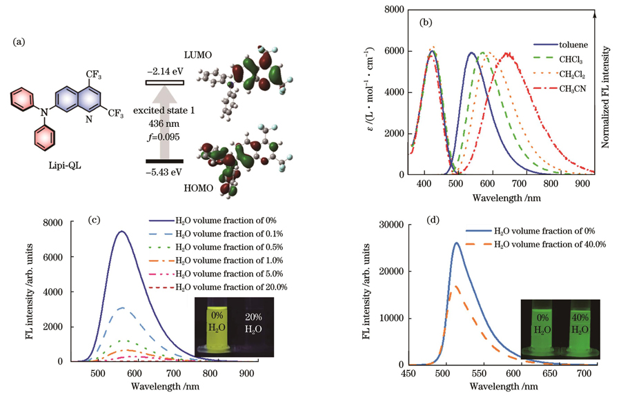
As shown in Fig.1(c), the probe exhibits highly efficient fluorescence emission when the water volume fraction is 0, indicating that it can exhibit high fluorescence intensity within lipid droplets. When the water volume fraction gradually increases, the probe exhibits extremely sensitive fluorescence quenching properties: quenching most of the fluorescence emission when the water volume fraction is only 1%. When the water volume fraction increases to 20%, the probe's emission is almost completely quenched, and the fluorescence signal disappears. This indicates that even if a small portion of the probe enters the cell and stains organelles other than lipid droplets, the fluorescence emission is quenched by the polar environment in which it is placed, thus showing a high selectivity for lipid droplet staining. We also test the fluorescence switching characteristics of the commercial lipid droplet dye, BODIPY 493/503. As shown in Fig.1(d), the fluorescence quenching of BODIPY 493/503 in the dioxane solution with 40% water volume fraction is not apparent, which may be the main reason for its poor lipid droplet staining selectivity. Figure 3 shows that the Lipi-QL fluorescent probe efficiently stains cellular lipid droplets at different concentrations. In contrast, BODIPY 493/503 stains lipid droplets much less selectively, staining other membrane-like cellular structures in addition to cellular lipid droplets with a lower imaging signal-to-noise ratio. This staining selectivity comparison highlights the significant advantage of the polar quenching luminescence property of the Lipi-QL fluorescent probe for the efficient and selective labeling of cellular lipid droplets. After washing the free probe with phosphate buffered saline (PBS), three-dimensional confocal imaging is performed. The experiment is performed at a high xy-plane point resolution with a small z-sweep step (200 nm) to obtain high-quality 3D confocal photographs (Fig.4). The spatial distribution of intracellular lipid droplets can be seen clearly in this photograph, demonstrating the usefulness of the probe for 3D confocal imaging. The Lipi-QL fluorescent probe is also used for multicolor confocal imaging because of its excellent performance. The nuclei, lipid droplets, lysosomes, and mitochondria of live HeLa cells are stained with the Hoechst 33342 commercial dye for nuclei, Lipi-QL commercial dye for lipid droplets, LysoTracker Deep Red commercial dye for lysosomes, and MitoTracker Deep Red commercial dye for mitochondria, respectively. High-quality four-color confocal images of living cells are successfully obtained by performing confocal fluorescence. Based on the different absorption and emission spectra of these four fluorescent probes, imaging is performed through line-by-line scanning, effectively avoiding the occurrence of crosstalk between individual fluorescent channels.
In conclusion, an advanced lipid droplet fluorescent probe with fluorescence switching properties, Lipi-QL, is developed in this study, which allows for the efficient and selective labeling of cellular lipid droplets. The probe also has high fluorescence brightness, a large Stokes shift, and good biocompatibility. Based on these excellent properties, high-quality three-dimensional confocal imaging of fixed cells and four-color confocal imaging of live cells are successfully achieved using this probe, highlighting its utility in lipid droplet fluorescence imaging. The development of this probe provides an effective tool for cell biology studies of lipid droplets and a new approach for the design and synthesis of highly labeled selective fluorescent probes.
1 引言
脂滴是一种球形细胞器,广泛存在于绝大部分的真核细胞中。其内部是由甘油三酯和胆固醇酯等构成的中性脂质核,核外包覆的是附着各种蛋白的磷脂单层膜[1-2]。脂滴从内质网的双层膜间以出芽的方式脱落进入细胞质中,所形成的新生脂滴尺寸较小,它们会在细胞质中多种酶的作用下逐渐成熟。在过去很长一段时间内,脂滴都被认为只是细胞中储存过量脂质的惰性脂肪颗粒。近年来研究发现,脂滴是细胞中一种重要的细胞器,参与能量代谢、膜转运和炎症反应等多种生理活动[3-5],并且脂滴的功能失调与多种代谢类疾病密切相关,如神经退行性疾病、肥胖症和癌症等[6-8]。因此,脂滴研究成为了近年来细胞生物学领域最热门的方向之一。
荧光成像[9-11]技术是观察脂滴并研究其生理功能最有力的工具之一。其中,共聚焦成像是目前使用最为广泛的荧光成像技术,相应地用于共聚焦成像具有高荧光亮度的脂滴荧光探针研究也有不少报道[11-23]。最为著名的当属已经商业化的Nile Red和BODIPY 493/503[13]。但这两种探针仍有一些缺点,例如两者的染色选择性都不够高,除了染色脂滴外,这两种荧光探针还会染色细胞中的其他结构。为了提升脂滴荧光探针的标记选择性,一种有效的方法是通过调节荧光探针的分子结构来改善其亲疏水性,从而显著提高荧光探针的脂滴染色特异性。例如,Collot等[12]报道了一系列基于单苯环骨架的红光发射脂滴荧光探针,通过调节分子结构中烷基链的长度和形状,改变荧光分子的亲疏水性和细胞膜通透性,得到了油水分配系数(CLogP)值在6左右的染色性能最佳的脂滴荧光探针Ph-Red。另外一种有效提升脂滴荧光探针标记选择性的方法是通过合理地设计具有荧光开关特性的荧光探针,实现低极性的脂滴内荧光点亮,高极性环境中荧光淬灭,从而实现高的脂滴染色特异性。例如,李晨梦等[9]开发了一系列具有荧光开关特性的高亮度脂滴荧光探针,由于这些探针在细胞质等极性环境下荧光会显著淬灭,从而在细胞和组织中都实现了高信噪比的脂滴荧光成像。这些工作显著地促进了脂滴的细胞生物学研究。但总体来说,能够高亮度和高效选择性标记细胞脂滴的荧光探针仍十分有限。
本文研制了一种极性敏感的具有荧光开关特性的脂滴荧光探针(Lipi-QL)。该探针分子结构简单,合成容易,具有给受体结构和显著的极性淬灭特性,还具有高的荧光亮度、高的脂滴染色选择性和低的细胞毒性。将该探针用于脂滴的共聚焦荧光成像,在不同的染色浓度下实现了显著高于商用探针BODIPY 493/503的成像信噪比。考虑到该探针不错的光稳定性,将该探针用于三维共聚焦成像,获得了高质量的能直观展现脂滴空间分布的三维图片。使用该探针搭配其他三种商用染料,成功实现了活细胞的四色共聚焦成像。这些应用突出了Lipi-QL高的成像信噪比,说明了该探针在脂滴荧光成像上的实用性。另外,该探针的分子设计为高效选择性标记细胞器的荧光探针研制提供了新的思路。
2 结果与讨论
2.1 荧光探针分子设计及光物理性质
Lipi-QL的分子结构是基于喹啉的分子骨架,在一端引入两个三氟甲基增强其吸电子能力,在另一端引入二苯胺给体。这样得到的给受体分子展现出了高荧光亮度,同时还具有大的斯托克斯位移,如

图 1. 荧光探针的设计、合成与光物理性质。(a)Lipi-QL的分子结构及其TD-DFT计算结果;(b)Lipi-QL在不同极性有机溶剂中的吸收发射光谱;(c)Lipi-QL在不同二氧六环溶液中的荧光光谱及荧光照片;(d)BODIPY 493/503在不同二氧六环溶液中的荧光光谱及荧光照片
Fig. 1. Design, synthesis, and photophysical properties of fluorescent probes. (a) Molecular structure of Lipi-QL and its calculation results by TD-DFT; (b) absorption emission spectra of Lipi-QL in different polar organic solvents; (c) fluorescence spectra and fluorescence photographs of Lipi-QL in different dioxane solutions; (d) fluorescence spectra and fluorescence photographs of BODIPY 493/503 in different dioxane solutions
之后研究了Lipi-QL在不同极性有机溶剂中的光物理性质[
2.2 荧光开关特性
由于极性淬灭发光特性对于提升荧光成像的信噪比具有重要意义,因此在这里详细地研究了Lipi-QL的荧光开光特性。细胞脂滴内的极性与甲苯或二氧六环溶剂的极性相当[18-19],因此Lipi-QL在细胞脂滴内的发光特性与其在甲苯或二氧六环中的类似。通过改变二氧六环溶液中水的含量来模拟脂滴以及其他细胞器中的极性环境。如
2.3 细胞脂滴染色共定位实验
通过3-(4,5-二甲基噻唑-2)-2,5-二苯基四氮唑溴盐(MTT)测试研究了Lipi-QL的生物相容性。在探针浓度为10 μmol/L的情况下孵育24 h后,没有观察到细胞毒性(详见支撑材料

图 2. HeLa细胞中Lipi-QL和Ph-Red的共定位实验(标尺为10 μm)。(a)Lipi-QL的绿色成像通道;(b)Ph-Red的红色成像通道;(c)两个荧光通道以及明场的叠加图片
Fig. 2. Co-localization experiments of Lipi-QL and Ph-Red in HeLa cells (scale bar is 10 μm). (a) Green imaging channel of Lipi-QL; (b) red imaging channel of Ph-Red; (c) superimposed images of two fluorescence channels and bright field
2.4 Lipi-QL和BODIPY 493/503的染色选择性对比
之后对比了具有荧光开关特性的Lipi-QL与脂滴荧光探针BODIPY 493/503在不同浓度下的染色选择性。活的HeLa细胞分别用不同浓度(1 μmol/L和10 μmol/L)Lipi-QL和BODIPY 493/503染色0.5 h,用Hank's平衡盐溶液(HBSS)洗去游离的荧光探针后再进行共聚焦成像。如

图 3. 不同浓度下Lipi-QL与BODIPY 493/503在HeLa细胞中的染色选择性对比(标尺为10 μm)
Fig. 3. Comparison of staining selectivity between Lipi-QL and BODIPY 493/503 in HeLa cells under different concentrations (scale bar is 10 μm)
2.5 固定细胞的三维共聚焦成像
三维共聚焦成像是观察亚细胞结构空间分布的一种强有力工具。通常对样品进行多重成像扫描,通过三维重构多张z扫切片,从而得到三维图片,这些操作会对荧光探针造成严重的光漂白。所以具有高亮度和高光稳定性的荧光探针是三维成像迫切需求的。此外,非特异性染色照片的堆叠会使三维共聚焦图片变得模糊不清[18],因此高的标记选择性对于三维共聚焦成像也是格外重要的。而Lipi-QL刚好满足上述要求,所以将其用于三维共聚焦成像。利用商用显微镜上配备的LAS X软件对z扫切片进行3D重构,得到3D共聚焦图片。活细胞中脂滴的快速移动会显著降低三维成像的分辨率,因此在本实验中,活HeLa细胞用浓度为2 μmol/L的Lipi-QL染色2 h后,利用质量分数为4%的多聚甲醛在室温下固定15 min。用磷酸盐缓冲液(PBS)洗去游离探针后,进行三维共聚焦成像。该实验是在高的xy平面点分辨率以及小的z扫步幅(200 nm)下完成的,因此获得了高质量的三维共聚焦照片(

图 4. 使用Lipi-QL染色的HeLa细胞的三维共聚焦成像
Fig. 4. 3D confocal imaging of HeLa cells stained with Lipi-QL
2.6 活细胞多色共聚焦成像
多色共聚焦成像能够在一张图片上揭示不同的细胞活动过程,因此能够提供重要的生物学信息。基于Lipi-QL优异的性能,将其用于多色共聚焦成像。在本实验中,活HeLa细胞的细胞核、脂滴、溶酶体和线粒体分别用细胞核商用染料Hoechst 33342、脂滴荧光探针Lipi-QL、溶酶体商用染料Lyso Tracker Red和线粒体商用染料Mito Tracker Deep Red染色。之后进行共聚焦荧光成像,条件如下:Hoechst 33342的激发光波长为λex=405 nm,检测区间为λem=415~445 nm;Lipi-QL的激发光波长为λex=458 nm,检测区间为λem=470~550 nm;Lyso Tracker Red的激发光波长为λex=570 nm,检测区间为λem=580~610 nm;Mito Tracker Deep Red的激发光波长为λex=640 nm,检测区间为λem=650~750 nm。基于这四种荧光探针吸收和发射光谱的不同,通过线线逐序扫描的方式进行成像,有效避免了各个荧光通道之间串色的发生。因此,成功得到了高质量的活细胞四色共聚焦成像图片,如

图 5. 活Hela细胞的多色共聚焦成像(标尺为10 μm)。(a)Hoechst 33342染色的细胞核;(b)Lipi-QL染色的脂滴;(c)Lyso Tracker Red染色的溶酶体;(d)Mito Tracker Deep Red染色的线粒体;(e)四个荧光通道的合并图片
Fig. 5. Multicolor confocal imaging of live Hela cells ( scale bar is 10 μm). (a) Hoechst 33342-stained nucleus; (b) Lipi-QL-stained lipid droplets; (c) Lyso Tracker Red-stained lysosomes; (d) Mito Tracker Deep Red-stained mitochondria; (e) merged images of four fluorescence channels
3 结论
研制了一种先进的具有荧光开关特性的脂滴荧光探针Lipi-QL。该探针极性淬灭的发光特性使得其可以高效选择性地标记细胞脂滴。该探针还具有高的荧光亮度、大的斯托克斯位移和良好的生物相容性。基于这些优异的性能,使用该探针成功实现了高质量的固定细胞三维共聚焦成像和活细胞四色共聚焦成像,突出了该探针在脂滴荧光成像中的实用性。该探针的研制为脂滴的细胞生物学研究提供了有效工具,为高标记选择性荧光探针的设计合成提供了新的思路。
备注:本文支撑材料可扫下方二维码获得。
[1] Olzmann J A, Carvalho P. Dynamics and functions of lipid droplets[J]. Nature Reviews Molecular Cell Biology, 2019, 20(3): 137-155.
[2] Thiam A R, Beller M. The why, when and how of lipid droplet diversity[J]. Journal of Cell Science, 2017: 315-324.
[3] Thiam A R, Farese Jr R V, Walther T C. The biophysics and cell biology of lipid droplets[J]. Nature Reviews Molecular Cell Biology, 2013, 14(12): 775-786.
[4] Walther T C, Farese R V. Lipid droplets and cellular lipid metabolism[J]. Annual Review of Biochemistry, 2012, 81: 687-714.
[5] Farese R V,, Walther T C. Lipid droplets finally get a little R-E-S-P-E-C-T[J]. Cell, 2009, 139(5): 855-860.
[6] Roitenberg N, Cohen E. Lipid assemblies at the crossroads of aging, proteostasis, and neurodegeneration[J]. Trends in Cell Biology, 2019, 29(12): 954-963.
[7] Krahmer N, Farese R V,, Walther T C. Balancing the fat: lipid droplets and human disease[J]. EMBO Molecular Medicine, 2013, 5(7): 973-983.
[8] Liu Q P, Luo Q, Halim A, et al. Targeting lipid metabolism of cancer cells: a promising therapeutic strategy for cancer[J]. Cancer Letters, 2017, 401: 39-45.
[9] 李晨梦, 邵鹏飞, 吴柄萱, 等. 用于成像性能测试的荧光发光模拟系统[J]. 中国激光, 2022, 29(24): 2407204.
Li C M, Shao P F, Wu B X, et al. Fluorescence luminescence simulation system for imaging performance testing[J]. Chinese Journal of Lasers, 2022, 29(24): 2407204.
[10] 周陈娟, 潘文良, 陈同生. 青蒿琥酯诱导活性氧依赖性的细胞凋亡[J]. 中国激光, 2011, 38(2): 0204003.
[11] 邓大伟, 刘飞, 曹洁, 等. 两种近红外荧光探针的合成及肿瘤靶向研究[J]. 中国激光, 2010, 37(11): 2735-2742.
[12] Collot M, Fam T K, Ashokkumar P, et al. Ultrabright and fluorogenic probes for multicolor imaging and tracking of lipid droplets in cells and tissues[J]. Journal of the American Chemical Society, 2018, 140(16): 5401-5411.
[13] Guo L F, Tian M G, Zhang Z Y, et al. Simultaneous two-color visualization of lipid droplets and endoplasmic reticulum and their interplay by single fluorescent probes in lambda mode[J]. Journal of the American Chemical Society, 2021, 143(8): 3169-3179.
[14] Shi L, Li K, Li L L, et al. Novel easily available purine-based AIEgens with colour tunability and applications in lipid droplet imaging[J]. Chemical Science, 2018, 9(48): 8969-8974.
[15] Zhou R, Cui Y Y, Dai J N, et al. A red‐emissive fluorescent probe with a compact single‐benzene‐based skeleton for cell imaging of lipid droplets[J]. Advanced Optical Materials, 2020, 8(13): 1902123.
[16] Fam T, Klymchenko A, Collot M. Recent advances in fluorescent probes for lipid droplets[J]. Materials, 2018, 11(9): 1768.
[17] Collot M, Bou S, Fam T K, et al. Probing polarity and heterogeneity of lipid droplets in live cells using a push-pull fluorophore[J]. Analytical Chemistry, 2019, 91(3): 1928-1935.
[18] Ashoka A, Ashokkumar P, Kovtun Y P, et al. Solvatochromic near-infrared probe for polarity mapping of biomembranes and lipid droplets in cells under stress[J]. The Journal of Physical Chemistry Letters, 2019, 10(10): 2414-2421.
[19] Peng G S, Dai J N, Zhou R, et al. Highly efficient red/NIR-emissive fluorescent probe with polarity-sensitive character for visualizing cellular lipid droplets and determining their polarity[J]. Analytical Chemistry, 2022, 94(35): 12095-12102.
[20] Zhou R, Wang C G, Liang X S, et al. Stimulated emission depletion (STED) super-resolution imaging with an advanced organic fluorescent probe: visualizing the cellular lipid droplets at the unprecedented nanoscale resolution[J]. ACS Materials Letters, 2021, 3(5): 516-524.
[21] Liu G N, Peng G S, Dai J N, et al. STED nanoscopy imaging of cellular lipid droplets employing a superior organic fluorescent probe[J]. Analytical Chemistry, 2021, 93(44): 14784-14791.
[22] Liu G N, Dai J N, Zhou R, et al. A distyrylbenzene-based fluorescent probe with high photostability and large Stokes shift for STED nanoscopy imaging of cellular lipid droplets[J]. Sensors and Actuators B: Chemical, 2022, 353: 131000.
[23] Taki M, Kajiwara K, Yamaguchi E, et al. Fused thiophene-S, S-dioxide-based super-photostable fluorescent marker for lipid droplets[J]. ACS Materials Letters, 2021, 3(1): 42-49.
Article Outline
赵力, 周日, 刘冠男, 彭桂衫, 王晨光, 贾晓腾, 卢革宇. 具有荧光开关特性的喹啉衍生物用于高效选择性标记细胞脂滴[J]. 中国激光, 2023, 50(9): 0907102. Li Zhao, Ri Zhou, Guannan Liu, Guishan Peng, Chenguang Wang, Xiaoteng Jia, Geyu Lu. Quinoline Derivatives with Fluorescent Switching Properties for Efficient Selective Labeling of Cellular Lipid Droplets[J]. Chinese Journal of Lasers, 2023, 50(9): 0907102.


