眼屈光度与生物参数测量的一体化设计与实现
Myopia is a common eye disease that has reached an epidemic level of 90% among university students in China. It has emerged as the leading cause of vision loss in Chinese adolescents. Myopia prevention and control have become a social concern and a national strategy. Establishing refractive development files and predicting refractive error based on these data is one of the most efficient ways to prevent and control myopia. There are many ocular biometric measuring instruments and optometers in the market, but there are no devices dedicated to the data acquisition of refractive development files. Based on previous research, this study combined interference, wavefront aberration, and image analysis to establish an integrated measurement system of ocular refraction and biological parameters, mainly measuring parameters including the axial length (AL), corneal curvature (K), anterior chamber depth (ACD), and diopter. We hope to provide an integrated, automated, and intelligent data collection, storage, and analysis platform for the refractive development of myopia prevention and control.
An integrated system for the measurement of the ocular refractive error and ocular biometry consists of a Twyman-Green low-coherence interference subsystem and a Shack-Hartmann wavefront sensing subsystem (Fig.1). A super-luminescent diode (SLD), beam splitter (BS), prism retroreflector, polarization beam splitting (PBS), and avalanche photodiode (APD) compose the Twyman-Green low-coherence interference subsystem. The SLD of the Twyman-Green low-coherence interference subsystem, lens relaxed optical path, and Shack-Hartmann wavefront sensor form the ocular refractive error-measuring optical subsystem. The axial length of the human eye was measured based on the Twyman-Green low-coherence interference system. Corneal reflex imaging was used to measure the corneal curvature. Measurement of anterior chamber depth is achieved in combination with slit lamp and corneal curvature measurement. The integrated design was applied in the optical system design and fused wavefront aberration analysis and interference optical paths; the strict collimation SLD could be a shared source to achieve wavefront aberration analysis and eye-axis measurement. Diopter measurements were calibrated using standard model eyes to ensure the accuracy of the measurement results. The biological parameters of the human eye (AL, K, and ACD) and diopter (spherical and cylindrical degrees) were measured five times for each subject using the experimental prototype and IOL Master 500, respectively. All statistical analysis were performed using SPSS 25.0 software. The repeatability of the experimental prototype measurements was evaluated using the within-subject standard deviation(Sw), test-retest repeatability (TRT), coefficient of variation (CoV), and intra-group correlation coefficient (ICC). The measurement results of the experimental prototype and IOL Master 500 were compared to analyze their accuracy and consistency.
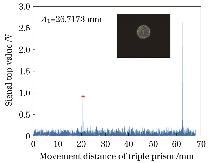
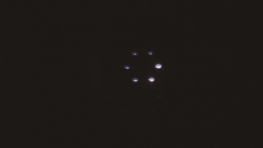
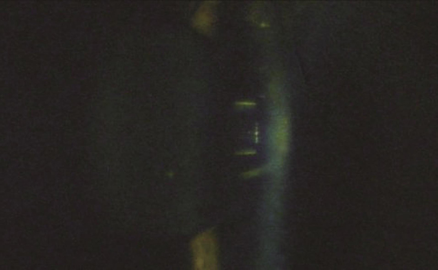
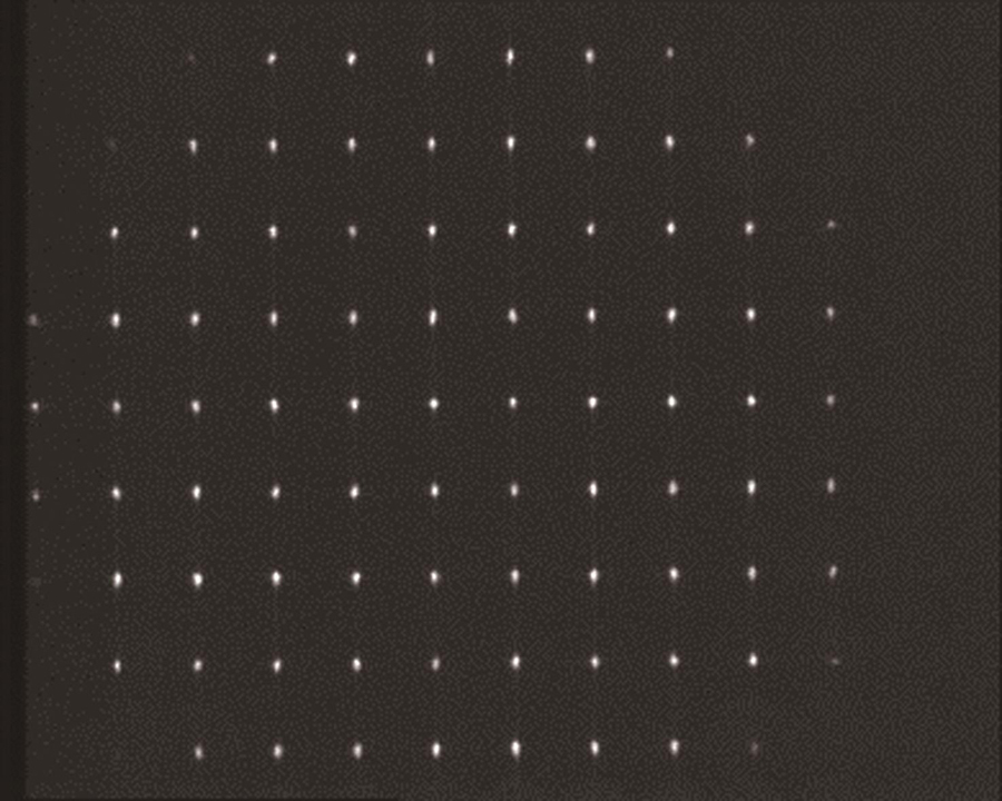
Five volunteers were recruited inside the laboratory, and the operator measured the subject’s left eye using the system experimental prototype and randomly showed the result of the eye axis length, radius of curvature, anterior chamber depth, and images of the measured human eye on the wavefront aberration sensor (Fig.3, Fig.4, Fig.5, Fig.6). The quality of the acquired images was good. The experimental prototype was used to measure the standard eye model with different diopters. The results of the single-sample t-test showed that the significance of the P value was greater than 0.05 (Table 1), and there was no difference between the diopter detection value of the experimental prototype and the standard value of the standard eye model, indicating that the experimental prototype could ensure measurement accuracy. From the values of Sw and TRT, it can be deduced that both the ocular biological parameters and diopter measurements have high repeatability. The CoV of eye biological parameters was less than 3%, the variation in the axial length and corneal curvature was smaller (CoV is less than 0.256%), and the ICC was higher than 0.6, reflecting high consistency (Table 2). The Bland-Altman analysis of the eye axis length, radius of curvature, and anterior chamber depth measured by the experimental prototype and IOL Master 500 showed no significant difference (P>0.05); the difference range was very small, and the 95% consistency limit was -0.045-0.056 mm, -0.037-0.056 mm, -0.534-0.355 mm, respectively, and all the measurement results were within the consistency interval (Table 3).
In this study, we designed an ocular refractive and biometric measurement system based on the Hartmann sensor and low-coherence heterodyne interference. Following previous studies, a standard eye model was used as the calibration sample to observe the diopter measurement results of the system. From the result analysis, it can be concluded that the results of this prototype measurement were highly consistent with the IOL Master 500 measurement results, and there was no significant difference in the results. The results showed excellent consistency of the ocular biological parameters and refractive measurements obtained using the integrated experimental prototype and IOL Mater 500. With the development of technology, it will be possible to provide special refractive file data acquisition for the prevention and control of myopia. In the future, we hope to provide an integrated, automated, and intelligent data collection, storage, and data analysis platform for refractive developmental files for myopia prevention and control.
1 引言
目前全球近视患者与日俱增,已成为全球关注的严重的公共卫生问题之一[1]。低龄化近视的进展可导致高度近视,从而增加近视致盲性并发症的风险,如视网膜脱离、近视性黄斑变性和青光眼等[2-3]。国家卫生健康委员会于2020年发布的《中国眼健康白皮书》显示,2018年全国儿童青少年总体近视率为53.6%,其中6岁儿童的近视率为14.5%,小学生为36%,初中生为71.6%,高中生为81%[4]。近视已成为我国儿童和青少年视力下降的主要原因。现有的近视防控方法包括健康用眼、药物治疗、手术治疗、配戴角膜塑形镜或近视防控框架眼镜。随着人们对近视防控意识的逐渐深入,各种防控策略都占有一定的市场地位,但目前还没有普适的防控策略。近视防控的关键在于早期预防,因为近视本身是不可治愈的,预防近视的发生与进展是关键。儿童和青少年的眼球处于快速发育阶段,并在发育过程中由远视状态向正视状态发育,因此,在不同的生长时期,眼球的状态不同。从横、纵断面的研究结果来看[5-11],学龄儿童眼生物参数随着年龄的增长而变化:等效球镜度降低,眼轴变长,晶状体变薄,前房深度增大,中央角膜变厚,轴率比(眼轴长与角膜曲率的比值)增大,但角膜曲率半径无明显变化。这些研究结果表明,在儿童发育过程中,眼生物参数的变化对屈光发育起到了至关重要的作用。眼生物参数主要包括眼轴长度(AL)、角膜曲率(K),前房深度(ACD)。《2022年中国儿童青少年视觉健康白皮书》中提到,建立屈光发育档案,通过定期记录孩子的身高、体重、裸眼视力、眼轴长度、角膜曲率、前房深度、眼压、屈光状态等与视力发展相关的项目,可以密切跟踪孩子的视觉健康发育情况。建立屈光发育档案是防治近视的基础性工作,目的是充分了解近视眼发生发展的规律,寻找确实有效的、科学的防治措施。目前市面上的眼科主流仪器都具有其独有的技术方案,要么是眼生物参数测量仪,要么是屈光度测量仪。国内很多研究团队在测量相关数据方面进行了大量工作,如:中山大学中山眼科中心林浩天教授团队[12]首次将深度学习应用于基于眼部图像的近视筛查,并实现了较高的筛查精度,能够远程监测近视儿童眼球的屈光状态;刘春华等[13]设计了一种偏心摄影视力筛查设备的成像光学系统,该系统可以减小偏心摄影验光设备测量人眼屈光度的误差,使拍摄的人眼照片更加清晰;陈春璐等[14]提出了一种基于噪声模型变换的子孔径光斑质心提取方法,该方法可以充分提升子孔径光斑的质心定位精度和稳定性。复旦大学附属眼耳鼻喉科医院和上海眼病防治中心都在屈光档案方面做了非常多的工作[15-18]。笔者检索发现有相关研究人员申报了眼生物参数与屈光度测量的一体化设计专利,该技术是基于光学相干层析成像(OCT)结合夏克-哈特曼波前相差分析技术实现的[19]。为了保证眼生物参数、屈光度数据的可靠性和准确性,需要采用一体化设备完成数据的测量。
近年来,本团队在主客观人眼波像差分析[20]、眼生物参数测量[21-22]等方面进行了大量的基础和应用性研究工作:实现了主客观人眼波像差分析系统实验样机以及眼生物参数测量系统的工程样机[20];将ZEISS标准模拟眼作为标定样品,分析了系统的眼轴长度测量结果[21];将样机与IOL Master对模拟眼的眼轴测量结果进行了比较[22]。近期,本团队继续在眼屈光度测量方面开展工作,并进行了眼屈光度与眼生物参数的一体化测量设备的研发工作,旨在为屈光发育档案的数据采集和数据处理提供一体化、自动化和智能化数据采集、存储和分析利用平台。本文主要报道眼屈光度与生物参数测量的一体化设计与实现。
2 基本设计原理
2.1 整体设计思路
在前期的研究中,本团队建立了眼的波像差分析系统和眼生物参数测量系统。前者主要通过夏克-哈特曼(Shack-Hartmann)波前传感器实现人眼主客观波像差(包括球差和像散特性)分析[20];后者基于泰曼-格林(Twyman-Green)低相干干涉系统测量人眼的眼轴长度[21-22],采用角膜反射成像技术实现角膜曲率的测量,利用裂隙灯与角膜曲率测量实现前房深度的测量。基于上述基础,融合波像差分析系统和干涉系统,经过严格准直激光,可以共用同一个光源实现波像差和眼轴测量。具体的光路设计如

图 1. 眼屈光度与生物参数测量一体化设计光路图
Fig. 1. Optical path diagram with integrated design of eye refraction and biological parameter measurement
2.2 眼轴长度测量光路设计
本团队设计的眼轴长度测量系统[21](New AL)是基于双光路外差干涉原理搭建的泰曼-格林干涉系统,该系统主要由近红外超辐射发光二极管S3(中心波长为790 nm,带宽为30 nm,偏振度为1000∶1)、准直镜L14、分光棱镜BS(分束比为50∶50)、角锥棱镜P1和P2、光电探测器D和数据采集卡(DAQ)组成。干涉信号经光电探测器转换为电信号后再由前置放大器Pre放大,之后被数据采集卡采集并被输入计算机,对数据进行分析即可得到眼轴长度。
2.3 角膜曲率测量光路设计
角膜曲率测量光路的设计原理是角膜反射成像。角膜曲率测量方法为:6个呈正六边形排布的LED光源(S21、S22等)经过1 mm孔径光阑(I21、I22等)后变为点光源,点光源经透镜(L21、L22等)准直后投射到角膜前表面,角膜反射的图像经L7、L8及L10~L13组成的物方远心光路后成像在CCD2上;利用重心算法求出角膜反射图像中位于正六边形长对角线上的两个点光源的距离,便可求得角膜曲率。
2.4 前房深度测量光路设计
LED光源S1发出的光经光阑I1、柱面镜L1形成裂隙光,裂隙光经眼睛的晶体和角膜反射后进入L7、L8及L10~L13组成的物方远心光路并成像在CCD2上,如
式中:ACD为前房深度;R为角膜曲率半径;x为角膜与晶体反射像的间距;n为角膜折射率,一般取1.336;ω为裂隙灯入射角。
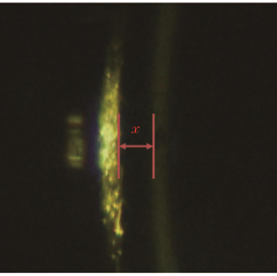
图 2. 角膜和晶体反射后所成的像及距离x测量
Fig. 2. Image formed by reflection of cornea and lens and distance x measurement
2.5 屈光度测量光路设计
在本设计中,采用准直的SLD(S3)激光作为参考波前的光源,其由眼轴测量光路进入人眼,经眼底反射后携带人眼的光学波前,经BS2、BS1以及透镜L5和L6组成的望远系统进入夏克-哈特曼波前传感器。其中,透镜L5、L6组成的望远系统将眼底与传感器成像相面形成光学共轭结构。同时,LED光源S4照明视标P图后经L3、R1、L4、BS2、BS1进入人眼,通过视标P的运动来调节屈光度误差,配合夏克-哈特曼波前传感器实现人眼波前的图像采集。通过图像处理计算出透镜阵列光斑的移动量,使用最小二乘法进行波前重建。波前像差使用7阶36项Zernike多项式展开,根据Zernike系数与球镜度、柱镜度的关系计算临床上的球差、散光及散光轴位数值[20]。
3 实验及结果
在实验室内招募了5名志愿者,因为都是年轻人群,因此没有设置排除条件。虽然这5名志愿者的眼球有不同的屈光状态,但基本都是健康的。采用本团队搭建的实验样机对5位受试者的左眼进行测量,采集的典型结果如
4 人眼测量结果统计
屈光度测量采用中国计量科学研究院校准且溯源的标准模拟眼进行标定,以确保测量结果的准确性,结果一致后再对实际人眼进行测量。采用实验样机测量0 D、±5 D、±10 D、±15 D,±20 D等9个屈光状态下的标准模拟眼的屈光度,测量9组数据,每组测量5次。对测量结果进行单样本t检验,以验证实验样机对标准模拟眼的测量重复性,测量结果分析如
测量人眼生物参数(眼轴长度、角膜曲率、前房深度)和屈光度(球镜度、柱镜度),每位受试者测量5次。使用组内标准差(Sw)、测试重复性(TRT)、变异系数(CoV)和组内相关系数(ICC)评估实验样机测量的可重复性,测量结果分析如
表 1. 标准模拟眼屈光度测量结果分析
Table 1. Analysis of diopter measurement of standard model eye
|
表 2. 人眼生物参数和屈光度测量结果分析
Table 2. Analysis of measured human eye biological parameters and diopter
|
表 3. IOL Master与实验样机的参数测量结果分析
Table 3. Analysis of parameter measurement results of IOL Master and experimental prototype
| ||||||||||||||||||||||
所有统计分析均用SPSS 25.0统计软件进行;采用Shapiro-Wilk正态分布进行检验,所有数据均符合正态分布特征。
5 结论与展望
结合光外差干涉和波像差分析等技术,设计了眼屈光度与生物参数测量的一体化系统,实现了眼轴长度、角膜曲率、前房深度和屈光度等参数的测量。一体化实现过程的难点主要在于眼轴长度的测量。测量眼轴长度需要获取角膜表面的干涉峰值信号与眼底视网膜干涉的峰值信号,而眼底视网膜信号较弱,干涉信号的信噪比不高。为了提高信噪比,在两方面进行了改进:一是提高电机速度的控制精度,降低信号频率范围,保证信号频率稳定,提高信号的提取能力;二是采用数字信号处理的方式,在不损失信号强度的前提下大大提高了信噪比。未来,也可以对眼轴信号进行多次测量,利用信号处理手段来提高信号的提取能力,进一步提高检出率。需要说明的是,本研究纳入的受试者均为年轻学生,不包括儿童和老年人,因此在评估不同健康水平和不同年龄段人群的眼屈光度时存在一定的局限性。从笔者获取的人眼的测量数据可以看出,所搭建的系统具有出色的重复性,尤其是在眼轴长度和角膜曲率测量方面具有很高的稳定性。虽然该系统在测量前房深度和屈光度方面仍然存在误差,但随着数据量的不断增加,可以通过一些技术手段来解决,比如对前房深度的提取算法进行优化。屈光度测量准确性的提高可以从以下两方面进行:
1)硬件方面。本次实验使用的基于夏克-哈特曼传感器的测量系统有动态范围、灵敏度、探测精度等指标,可从微透镜阵列尺寸、CCD尺寸、CCD分辨率和透镜焦距这几个参数中选择适合人眼屈光度测量的最优参数。
2)算法方面。基于夏克-哈特曼传感器的测量技术测量屈光度的主要步骤是图像预处理、质心计算、斜率计算、波前重构、屈光度计算,而基于夏克-哈特曼传感器的人眼像差检测算法的精度取决于波前重构的精度,由波前重构理论可知光斑阵列的质心探测精度决定了波前重构的精度。因此可以从质心计算入手,提出新算法或改进相关算法,进一步提高屈光度测量的重复精度。
在接下来的研究中,拟将迭代瞳孔跟踪和自动化运动控制技术应用于所设计的系统,以实现数据的采集自动化;然后结合大数据和人工智能实现智能化的眼屈光发育状态分析,最终使所提系统成为一套集眼屈光度和生物参数测量于一体的多参数一体化、智能化和个性化近视防控专用设备。
屈光度和眼生物参数是评价青少年身体素质的重要指标。目前,视力筛查的主要工具是验光仪,而眼生物参数的测量主要依靠眼生物参数测量仪。设计眼屈光度和生物参数一体化测量系统,不仅可以减少设备成本和设备操作所需的人力,还可以更加便捷快速地获取测量数据,建立屈光发育档案,比较眼生物参数和屈光度的变化,寻求青少年眼屈光发育的特点和规律,预测个体及群体的近视发展趋势,为早期近视干预控制提供科学依据和前瞻性指导。
[1] Morgan I G, Ohno-Matsui K, Saw S M. Myopia[J]. The Lancet, 2012, 379(9827): 1739-1748.
[2] Baird P N, Saw S M, Lanca C, et al. Myopia[J]. Nature Reviews Disease Primers, 2020, 6(1): 1-20.
[3] Saw S M, Gazzard G, Shih-Yen E C, et al. Myopia and associated pathological complications[J]. Ophthalmic & Physiological Optics, 2005, 25(5): 381-391.
[5] Deng L, Pang Y. Effect of outdoor activities in myopia control: meta-analysis of clinical studies[J]. Optometry and Vision Science, 2019, 96(4): 276-282.
[6] 朱田柱, 袁野, 周之晖, 等. 上海某社区青少年近视屈光不正筛查结果纵向分析[J]. 中国学校卫生, 2021, 42(6): 931-934.
Zhu T Z, Yuan Y, Zhou Z H, et al. Longitudinal analysis of myopia and refractive errors screening results in adolescents in a community in Shanghai[J]. Chinese Journal of School Health, 2021, 42(6): 931-934.
[7] 李龙, 陶佳, 范浩博, 等. 南充市城区学龄前儿童视力和屈光不正现状[J]. 川北医学院学报, 2020, 35(4): 612-615.
Li L, Tao J, Fan H B, et al. Current status of vision acuity and ametropia of preschool children in urban area of Nanchong[J]. Journal of North Sichuan Medical College, 2020, 35(4): 612-615.
[8] 张学辉, 艾欣, 李盼, 等. 3~12岁儿童屈光状态与眼部生物学参数的相关关系[J]. 国际眼科杂志, 2020, 20(8): 1409-1412.
Zhang X H, Ai X, Li P, et al. Relationship between refractive status and ocular biological parameters in children aged 3-12 years[J]. International Eye Science, 2020, 20(8): 1409-1412.
[9] Guo X X, Fu M, Ding X H, et al. Significant axial elongation with minimal change in refraction in 3- to 6-year-old Chinese preschoolers: the Shenzhen kindergarten eye study[J]. Ophthalmology, 2017, 124(12): 1826-1838.
[10] Zhang L L, He X G, Qu X M, et al. Refraction and ocular biometry of preschool children in Shanghai, China[J]. Journal of Ophthalmology, 2018, 2018: 5205946.
[11] Li T, Jiang B, Zhou X D. Axial length elongation in primary school-age children: a 3-year cohort study in Shanghai[J]. BMJ Open, 2019, 9(10): e029896.
[12] Yang Y H, Li R Y, Lin D R, et al. Automatic identification of myopia based on ocular appearance images using deep learning[J]. Annals of Translational Medicine, 2020, 8(11): 705.
[13] 刘春华, 赵楠, 柴松均, 等. 偏心摄影视力筛查设备光学系统设计[J]. 激光与光电子学进展, 2022, 59(13): 1322002.
[14] 陈春璐, 赵旺, 赵孟孟, 等. 基于噪声模型变换的子光斑质心提取方法[J]. 光学学报, 2023, 43(3): 0312005.
[15] 吴敏. 学龄儿童眼球生物学参数及屈光变化观察[D]. 天津: 天津医科大学, 2016.
WuM. Investigation of ocular biometry and refraction changes for the school-age children[D]. Tianjin: Tianjin Medical University, 2016.
[16] Ma Y Y, Zou H D, Lin S L, et al. Cohort study with 4-year follow-up of myopia and refractive parameters in primary schoolchildren in Baoshan District, Shanghai[J]. Clinical & Experimental Ophthalmology, 2018, 46(8): 861-872.
[17] 褚仁远, 瞿小妹. 建立儿童屈光发育档案是预防近视的基础步骤[J]. 中华眼科杂志, 2009, 45(7): 577-579.
Chu R Y, Qu X M. Setting the individual file of ocular refractive development of children is the primary procedure in the prevention of myopia[J]. Journal of Command and Control, 2009, 45(7): 577-579.
[18] 邹海东, 何鲜桂. 扎实推进中国儿童青少年屈光发育档案的建立[J]. 中华眼视光学与视觉科学杂志, 2015, 17(10): 581-584.
Zou H D, He X G. An archive for refractive development in children and adolescents[J]. Chinese Journal of Optometry Ophthalmology and Visual Science, 2015, 17(10): 581-584.
[19] 胡积烨, 黄铭斌, 张浩, 等. 一种验光仪和眼科生物多参数测量仪及测量方法: CN114159020A[P]. 2022-03-11.
HuJ Y, HuangM B, ZhangH, et al. Optometry unit, ophthalmology biological multi-parameter measuring instrument and measuring method: CN114159020A[P]. 2022-03-11.
[20] 张培茗, 陈家璧, 庄松林. 基于信息融合的人眼波前像差测量[J]. 中国激光, 2009, 36(10): 2571-2576.
[21] 王成, 周岐, 陈奕君, 等. 基于低相干干涉测量的眼轴长度测量系统研究[J]. 中国激光, 2022, 49(5): 0507106.
[22] Wang C, Huangfu S N, Zhou Q, et al. The measurement of ocular axial length in normal human eyes based on an improved Twyman-Green interferometer[J]. Journal of Biophotonics, 2021, 14(10): e202100021.
Article Outline
王成, 朱俊, 熊利能, 董建荣, 戴磊, 项华中, 陈明惠, 郑刚, 赵婕, 张大伟. 眼屈光度与生物参数测量的一体化设计与实现[J]. 中国激光, 2023, 50(9): 0907208. Cheng Wang, Jun Zhu, Lineng Xiong, Jianrong Dong, Lei Dai, Huazhong Xiang, Minghui Chen, Gang Zheng, Jie Zhao, Dawei Zhang. Integrated Design and Implementation of Ocular Diopter and Biological Parameters Measurement[J]. Chinese Journal of Lasers, 2023, 50(9): 0907208.
