High Power Laser Science and Engineering, 2017, 5 (2): 02000e10, Published Online: Jul. 26, 2018
An investigation progress toward Be-based ablator materials for the inertial confinement fusion
Figures & Tables
Fig. 1. Be–Cu capsule. (a) Optical microscope image, (b) SEM image, (c) micro-CT image, (d) polished cross-section morphology, (e) cross-section morphology, (f) Cu dopant distribution at cross-section.
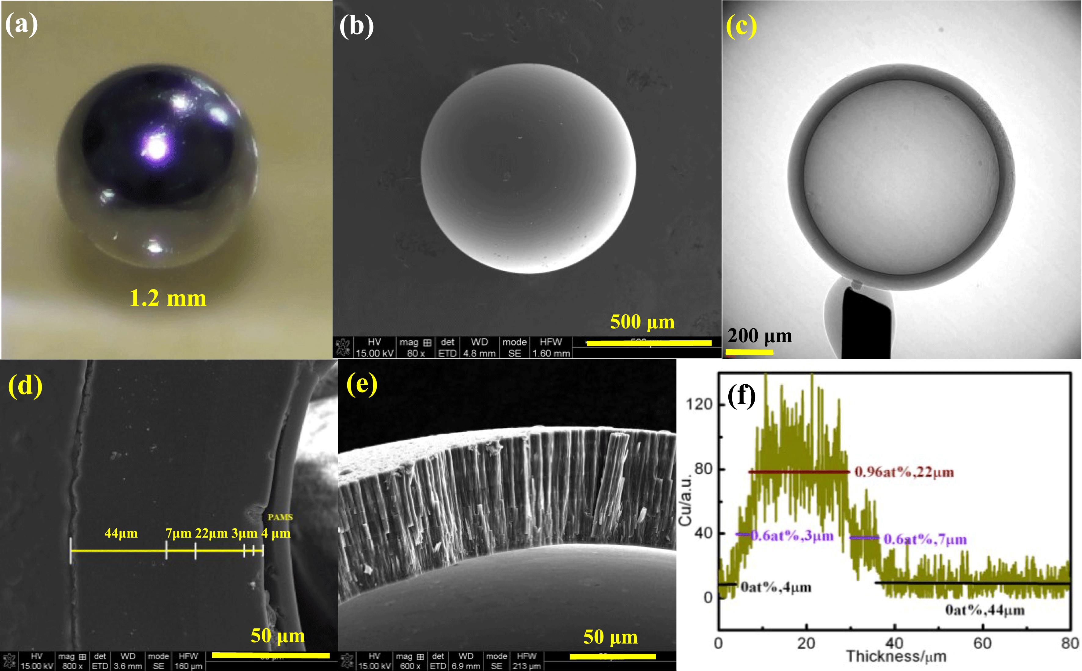
Fig. 2. Be coating morphologies. (a), (b) and (c) surface microstructure, cross-section microstructure and XRD pattern of Be coating prepared by thermal evaporation, (d), (e) and (f) surface microstructure, cross-section microstructure and XRD pattern of Be coating prepared by reactive evaporation.

Fig. 3. XPS spectra for films prepared at different $\text{CH}_{4}$ –Ar ratios after 30 min $\text{Ar}^{+}$ etching survey spectrum (a), deconvolution of Be1s peaks (b) and deconvolution of C1s peaks (c).

Fig. 4. (a) High-resolution TEM image and corresponding FFT pattern (inset upper right) for film deposited at room temperature, (b) typical XRD pattern of films with in situ annealing.

Fig. 5. Typical surface and cross-sectional morphologies of $\text{Be}_{2}\text{C}$ films with different thickness: (a) and (b) surface morphologies by AFM, (a1) and (b1) cross-sectional morphologies by SEM.

Fig. 6. Typical optical transmittance spectra of the $\text{Be}_{2}\text{C}$ films and corresponding photograph (inset).


Table1. The content of impurities in Be powder.
|
Bingchi Luo, Jiqiang Zhang, Yudan He, Long Chen, Jiangshan Luo, Kai Li, Weidong Wu. An investigation progress toward Be-based ablator materials for the inertial confinement fusion[J]. High Power Laser Science and Engineering, 2017, 5(2): 02000e10.


 PDF全文
PDF全文

