High Power Laser Science and Engineering, 2019, 7 (3): 03000e53, Published Online: Aug. 26, 2019
Single-shot electrons and protons time-resolved detection from high-intensity laser–solid matter interactions at SPARC_LAB  Download: 801次
Figures & Tables
Fig. 1. Experimental setup. The FLAME laser is sent to a stainless steel target. The charged particles emitted during this interaction are revealed by two single-shot time-resolved measurements: an electro-optical sampling diagnostic, able to measure the electric field carried by relativistic fast electrons, and a time-of-flight diamond detector, used to measure the temporal distribution of protons arriving on it and retrieve their energy spectra.
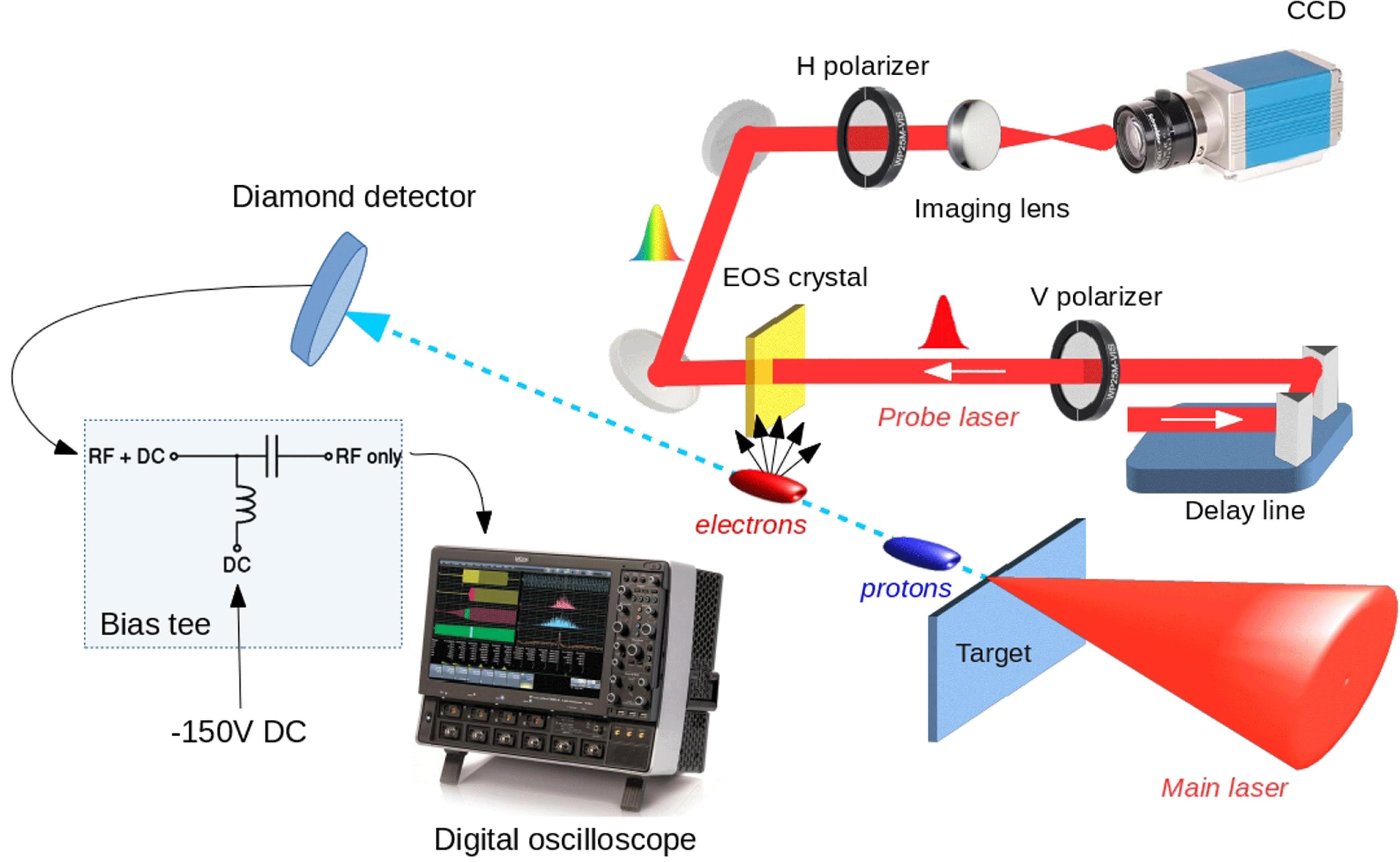
Fig. 2. Time-of-flight detector geometry. Schematic representation of the device layer structure (left) and picture of the surface Al interdigitated electrodes (right). The metal fingers were processed to $20~\unicode[STIX]{x03BC}\text{m}$ in width, with a spacing between the electrodes of $20~\unicode[STIX]{x03BC}\text{m}$ . The detector active area was approximately $2~\text{mm}^{2}$ .

Fig. 3. Typical 2D electric field carried by fast electrons as seen by our EOS diagnostic tool. The signal thickness is related to the temporal duration of the electric field. The typical shape is a direct consequence of our setup geometry[27].

Fig. 4. Line profile traced along the signal in Figure 3 . The measured electric field shows a peak value $E_{0}=1.5~\text{MV}/\text{m}$ and a temporal duration $\unicode[STIX]{x1D70F}=0.5$ ps FWHM. The fast electron charge also has been retrieved with a value of $Q=6$ nC.

Fig. 5. Typical signal provided by the TOF detector as seen by our 2 GHz Lecroy 620ZI oscilloscope. As one can see, it is possible to distinguish between two different signals arriving at different times: the first is associated with X-rays and low-energy electrons coming at the early stage of the interaction; the second is related to protons accelerated through the TNSA mechanism.

Fig. 6. Monte Carlo simulations by the SRIM code results for $10~\unicode[STIX]{x03BC}\text{m}$ of aluminium.


F. Bisesto, M. Galletti, M. P. Anania, M. Ferrario, R. Pompili, M. Botton, A. Zigler, F. Consoli, M. Salvadori, P. Andreoli, C. Verona. Single-shot electrons and protons time-resolved detection from high-intensity laser–solid matter interactions at SPARC_LAB[J]. High Power Laser Science and Engineering, 2019, 7(3): 03000e53.


 PDF全文
PDF全文

