激光与光电子学进展, 2018, 55 (5): 050003, 网络出版: 2018-09-11
线扫描共聚焦成像技术在生物医学成像中的应用  下载: 2368次
Application of Confocal Line Scanning Imaging Technique in Biomedical Imaging
图 & 表
图 1. 线扫描共聚焦成像系统结构示意图[5]
Fig. 1. Structure diagram of confocal line scanning imaging system[5]
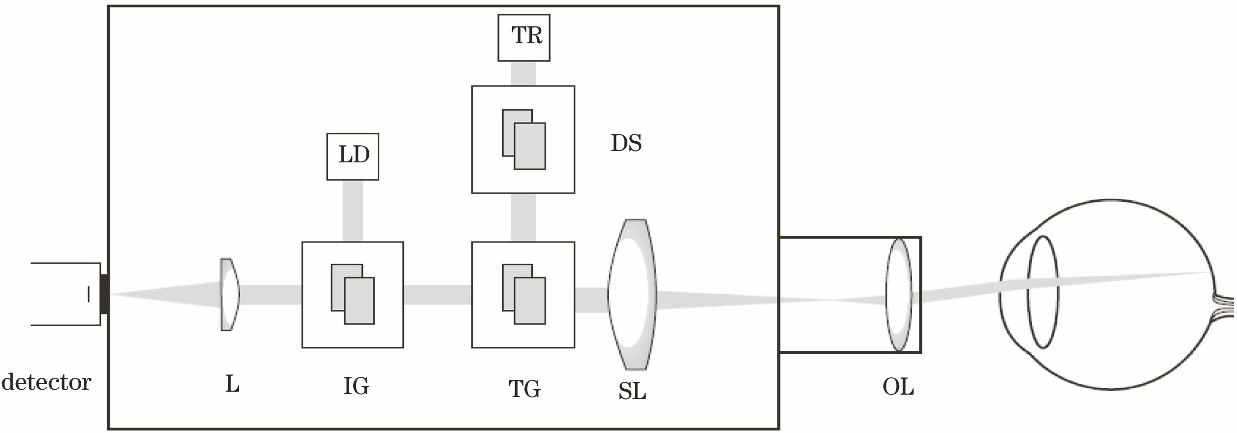
图 2. (a)横向分辨率[8]和(b)轴向分辨率的检测结果[8]
Fig. 2. Detection results of (a) lateral resolution[8] and (b) axial resolution[8]

图 3. (a)未消除抖动和(b)高帧频消抖后的视网膜图像[5]
Fig. 3. Fundus images (a) without shake cancellation and (b) with shake cancellation by using high frame frequency[5]

图 4. (a) 40 D和(b) 66 D屈光度眼底镜下的眼底血管图像[19]
Fig. 4. Fundus vessel images with (a) 40 D and (b) 66 D ophthalmoscopic lenses[19]

图 5. (a)以AOD为光偏转装置的成像系统结构及(b)横向分辨率检测结果[13]
Fig. 5. (a) Imaging system structure with AOD as light deflection device and (b) its lateral resolution detection result[13]

图 6. (a)线扫描共聚焦显微镜光路结构[8];(b)植物叶片细胞成像结果[8]
Fig. 6. (a) Light path of confocal line scanning microscopy[8]; (b) imaging result of plant leaf cells[8]

图 7. (a)共聚焦θ线扫描显微镜示意图及(b)皮肤细胞的在体成像结果[21]
Fig. 7. (a) Schematic for confocal theta line scanning microscope and (b) in vivo imaging result of skin cells[21]

图 8. (a)双轴线扫描共聚焦系统结构[22];(b)横向分辨率检测结果[22];(c)手持式双轴共聚焦显微镜结构图[14]
Fig. 8. (a) Structure of dual-axial confocal line scanning system[22]; (b) detection result of lateral resolution[22]; (c) structure of handle dual-axis confocal microscope[14]

图 9. (a)自适应线扫描共聚焦眼底成像系统结构及其(b)成像结果[25]
Fig. 9. (a) Structure of adaptive confocal line scanning fundus imaging system and its (b) imaging result[25]


孔文, 高峰, 樊金宇, 史国华. 线扫描共聚焦成像技术在生物医学成像中的应用[J]. 激光与光电子学进展, 2018, 55(5): 050003. Wen Kong, Feng Gao, Jinyu Fan, Guohua Shi. Application of Confocal Line Scanning Imaging Technique in Biomedical Imaging[J]. Laser & Optoelectronics Progress, 2018, 55(5): 050003.


 PDF全文
PDF全文

