光学学报, 2018, 38 (2): 0215005, 网络出版: 2018-08-30
基于机器学习的可降解支架检测与分割算法  下载: 1054次
Detection and Segmentation Algorithm for Bioresorbable Vascular Scaffolds Struts Based on Machine Learning
图 & 表
图 1. BVS支架贴壁分析流程图(第一幅图中的局部放大图显示了其中一个BVS支架的结构)
Fig. 1. Workflow of BVS strut malapposition analysis (Local enlarged drawing in the first image shows structure of one of the BVS struts)
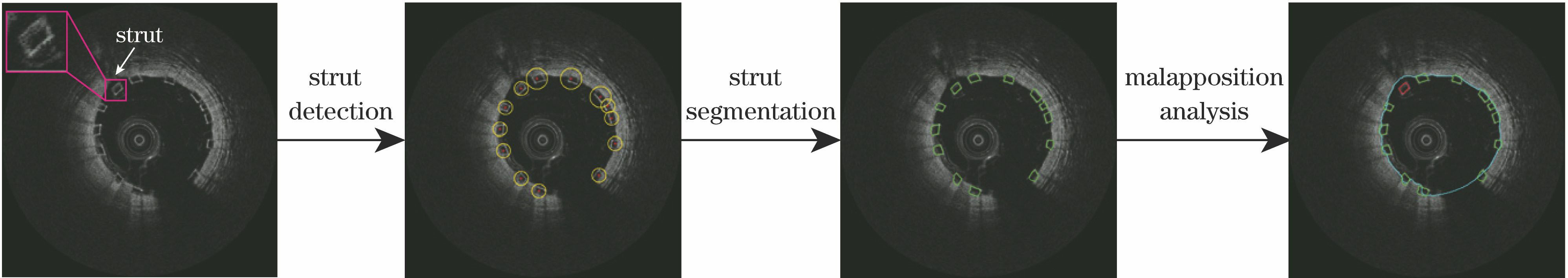
图 2. (a)单树桩弱分类器;(b)由图2(a)构建的强分类器;(c)三层决策树弱分类器;(d)由图2(c)构建的强分类器
Fig. 2. (a) Single stump-based weak classifier; (b) strong classifier boosted by Fig. 2(a); (c) three-layer decision tree-based weak classifier; (d) strong classifier boosted by Fig. 2(c)


图 4. 检测流程图。(a)输入图像;(b)检测区域;(c)滑窗示意图;(d)用级联分类器检测;(e)BVS候选点;(f)输出图像
Fig. 4. Workflow of detection. (a) Input image; (b) detection region; (c) diagram of sliding sub-window; (d) detection through cascaded classifier; (e) BVS candidates; (f) output image

图 5. 支架分割流程图。(a)笛卡尔坐标系下的一个支架;(b)极坐标系下的一个支架;(c)极坐标下的轮廓分割结果;(d)转回至笛卡尔坐标系下的分割结果
Fig. 5. Procedure of strut segmentation. (a) Strut in Cartesian coordinate system; (b) strut in polar coordinate system; (c) segmented contour in polar coordinate system; (d) segmented contour transformed back into Cartesian coordinate system

图 6. 支架贴壁分析结果图。(a)普通IVOCT图像;(b)(c)有血液伪影的图像;(d)~(f)同时包含贴壁良好和贴壁不良支架(对于贴壁不良支架,以白色线段表征其到血管壁的距离)
Fig. 6. Results of strut malapposition analysis. (a) Normal IVOCT images; (b)(c) images with blood artifacts; (d)-(f) images with both apposed and malapposed struts (For malapposed struts, distances between strut and lumen are represented by white lines)


表 1支架检测与分割结果
Table1. Results of strut detection and segmentation
| |||||||||||||||||||||||||||||||||||||||||||||||||||||||||||||||||||||||||||||||||||||
鲁逸峰, 金琴花, 荆晶, 陈韵岱, 曹一挥, 李嘉男, 朱锐. 基于机器学习的可降解支架检测与分割算法[J]. 光学学报, 2018, 38(2): 0215005. Yifeng Lu, Qinhua Jin, Jing Jing, Yundai Chen, Yihui Cao, Jianan Li, Rui Zhu. Detection and Segmentation Algorithm for Bioresorbable Vascular Scaffolds Struts Based on Machine Learning[J]. Acta Optica Sinica, 2018, 38(2): 0215005.


 PDF全文
PDF全文

