激光与光电子学进展, 2020, 57 (24): 240001, 网络出版: 2020-11-20
高速超分辨结构光照明显微的关键技术及应用  下载: 4166次封面文章特邀综述
High-Speed Structured Illumination Microscopy and Its Applications
图 & 表
图 1. SIM的重大理论和实验突破路线图[29-43]
Fig. 1. Roadmap of significant theoretical and technical breakthroughs of SIM[29-43]
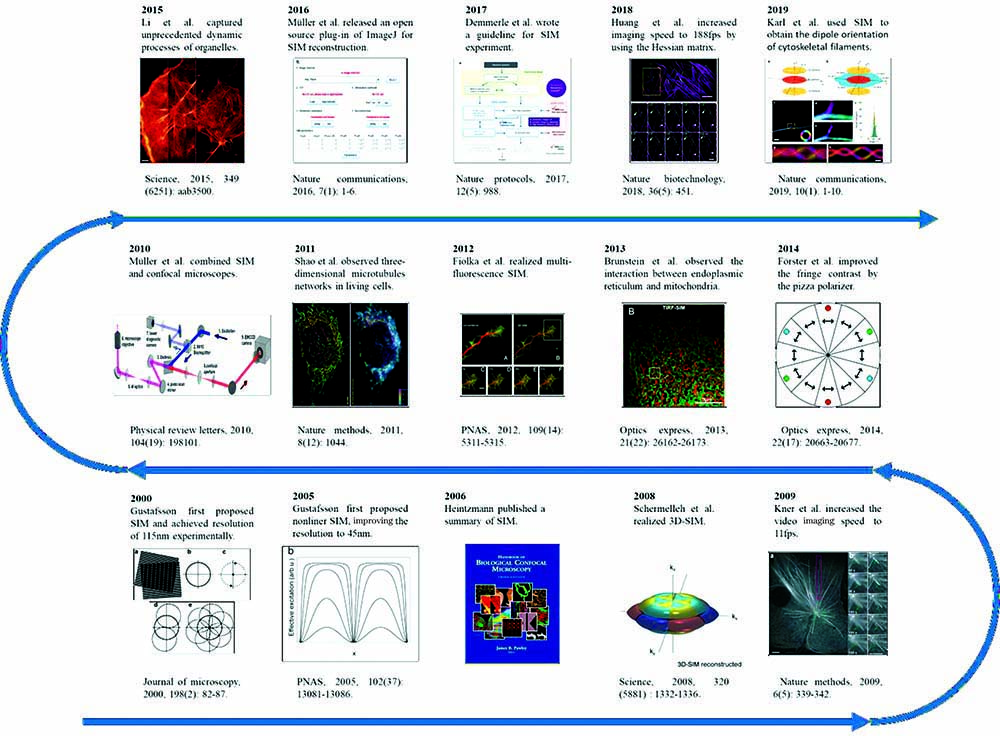
图 2. 莫尔条纹的产生。待测物和结构光叠加时会因拍频产生新的条纹[30]
Fig. 2. Generation of Moiré fringes. If sample structure is multiplied by structural light, beat pattern (Moiré fringes) will appear


图 4. 迭代法查找SLM加载光栅图样的流程图[56]
Fig. 4. Schematic diagram of iterative method for searching grating patterns displayed on SLM[56]

图 5. 光栅查找算法[56]。(a)像素化的光栅。白色和灰色分别表示SLM像素的开和关,红色为最小光栅单元起始点; (b)(c)两次2/3π相移时光栅(以紫色点为例)沿y方向移动的示意图
Fig. 5. Grating searching algorithm[56]. (a) Pixelated grating. White and gray represent on state and off state, respectively, and lattice points are pixels marked in red; (b)(c) grating with two steps of 2π/3 phase shift in y direction. Lattice point as a reference is marked in violet

图 6. Pizza偏振片和零级涡旋半波片。(a) Pizza偏振片[38];(b)零级涡旋半波片[61]
Fig. 6. Pizza polarizer and zero-order vortex half-wave retarder. (a) Pizza polarizer[38];(b) zero-order vortex half-wave retarder[61]


图 9. 三种初相位估计方法的结果对比[65]。(a)普通宽场反卷积的结果;(b)~(d)三种初相位估计算法的超分辨重图像;(e)~(h)局部放大结果。标尺:(a)~(d)为5μm;(e)~(h)为500nm
Fig. 9. Results of three initial phase estimation methods. (a) Deconvolution wide-field image; (b)--(d) super-resolution image reconstructed by three phase estimation algorithms; (e)--(h) magnified views. Scale bar: (a)--(d) 5μm; (e)--(h) 500nm

图 10. 密集肌动蛋白骨架结构的海森重构结果[42]。(a)宽场结果、传统维纳滤波结果和海森重构结果对比,标尺为2μm;(b)不同反卷积方法结果的局部放大图,标尺为500nm
Fig. 10. Densely packed cellular actin skeleton structures obtained by Hessian reconstruction[42]. (a) Wide-field image, traditional Wiener filtering result, and Hessian deconvolution result. Scale bar: 2μm; (b) magnified images reconstructed by different deconvolution methods. Scale bar: 500nm


图 12. 对鼠脑神经元形态功能的研究[75]。(a)以ChR2-GFP标记细胞膜,得到的宽场和SIM实验结果,标尺:5μm;(b)图12 (a)中宽场和SIM实验结果的OTF对比;(c)对细胞质进行标记的宽场和SIM实验结果,标尺:5μm;(d)图12 (c)中宽场和SIM的OTF对比;(e)按照时间先后顺序排列的SIM结果,展示了神经树突结构的动态变化,标尺:4μm
Fig. 12. Research on morphological function of mouse brain neurons[75]. (a) Deconvolved widefield and SIM images of cell membrane labeled as ChR2-GFP. Scale bar: 5μm; (b) OTFs of SIM and deconvolved widefield images in Fig. 12 (a); (c) deconvolved widefield and SIM images of labeled cytoplasm. Scale bar: 5μm; (d) OTFs of SIM and deconvolved widefield images in Fig. 12 (c); (e) time-

图 13. 商用SIM产品[83-85]。(a)德国蔡司公司Elyra 7;(b)日本尼康株式会社N-SIM;(c)美国通用电器公司GE DeltaVision OMX
Fig. 13. Commercial SIM products[83-85]. (a) Elyra 7 from Zeiss, Germany; (b) N-SIM from Nikon, Japan; (c) GE DeltaVision OMX from General Electric, America


图 15. SIM结合偏振信息确定蛋白质分子取向[43]。U2OS细胞微管蛋白的(a)宽场和(b)SIM结果对比,彩色为偏振方向,标尺:10μm;(c) SIM时间序列展示了微管运动的过程,标尺:1μm;(d)--(g)根据偏振信息建立的模型确定了α-tubulin分子取向
Fig. 15. Orientation of protein molecule determined by polarized information and SIM[43]. (a) Wide-field and (b) SIM images of microtubulin of U2OS cell. Scale bar: 10μm;(c) time-lapse SIM images showing the dynamic process. Scale bar: 1μm;(d)--(g) orientations of α-tubulin determined by model based on polarized information

表 1商用SIM产品部分参数对比
Table1. Partial parameters of commercial SIM products
|
赵天宇, 汪召军, 冯坤, 梁言生, 何旻儒, 云雪, 雷铭. 高速超分辨结构光照明显微的关键技术及应用[J]. 激光与光电子学进展, 2020, 57(24): 240001. Tianyu Zhao, Zhaojun Wang, Kun Feng, Yansheng Liang, Minru He, Xue Yun, Ming Lei. High-Speed Structured Illumination Microscopy and Its Applications[J]. Laser & Optoelectronics Progress, 2020, 57(24): 240001.


 PDF全文
PDF全文

