中国激光, 2020, 47 (2): 0207013, 网络出版: 2020-02-21
用于内窥光学相干层析成像探头的小型化及焦深拓展技术  下载: 2289次特邀综述
Probes for Endoscopic Optical Coherence Tomography: Minimized Design and Depth of Focus Extension
图 & 表
图 1. 探头原理图。(a)传统探头;(b)不含聚焦元件的多模干涉探头;(c)包含聚焦元件的多模干涉探头
Fig. 1. Schematic of probes. (a) Conventional probe; (b) multi-mode interference probe without focusing elements ; (c) multi-mode interference probe with focusing elements
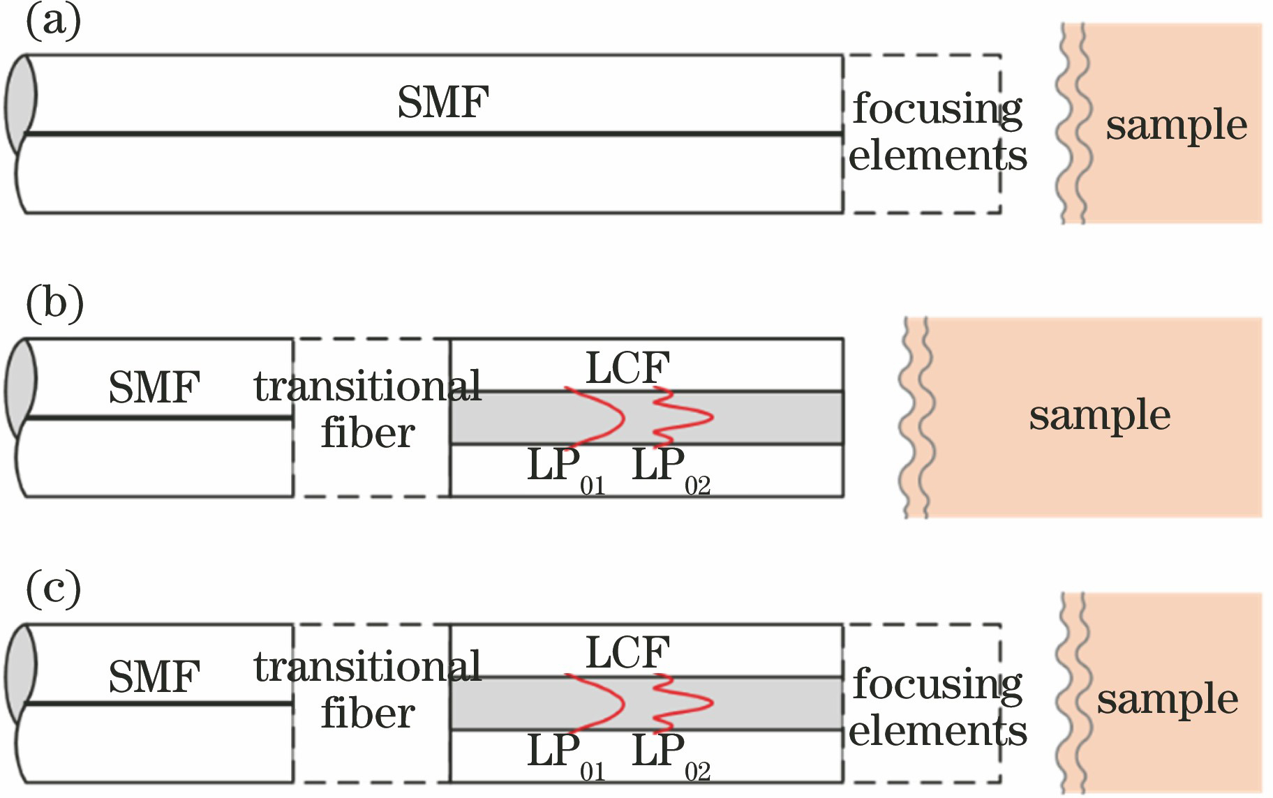
图 2. 探头原理图。(a)血管内OCT探头[23];(b)针式探头[24]
Fig. 2. Schematic of probes. (a) Intravascular OCT probe[23]; (b) imaging needle for OCT[24]

图 3. 基于球透镜的小型探头。(a)用于反射和聚焦光束的玻璃半球[25];(b)直径为500 μm的蓝宝石球透镜 [26];(c)能够同时用在空气中和液体中的直径为70 μm的共路OCT探头[27];(d)探头的光束大小随距探头末端距离的变化曲线[27]
Fig. 3. Miniature probe based on ball lens. (a)Glass hemisphere for reflecting and focusing beam[25]; (b) sapphire ball lens with 500 μm[26]; (c) 70 μm diameter probe for common-path OCT in air and liquids[27]; (d) curve of beam size of the probe changing with the distance from the end of the probe[27]

图 4. 光纤透镜的制作及其出射光斑。(a)~(c)光纤透镜的弧度半径随无芯光纤直径的增大而增大[28];(d)光纤透镜的弧度半径随电弧功率的增大而减小[29];(e)基于侧向抛光光纤透镜的侧向探头[30];(f)不经过保护套管时光斑的横向强度分布;(g)经过保护套管后的光斑横向强度分布[30]
Fig. 4. Fabrication of lensed fibers and their output beam spots. (a)-(c) Radii of the fiber lens increase with the diameters of the coreless fibers[28]; (d) radii of the fiber lens decrease with arc power of the splicer[29]; (e) side viewing probe design with angle polishing fiber lens[30]; (f) transverse intensity distribution of the spot without the pro

图 5. 超小型侧向针式探头[32-33]。(a)探头原理图;(b)使用针式探头获得的羊肺的三维OCT成像图;(c)使用针式探头获得的骨骼肌的OCT成像图;(d)骨骼肌的显微图
Fig. 5. Subminiature lateral probe[32-33]. (a) Schematic of probe; (b) three dimensional OCT image of sheep lung obtained by needle probe; (c) OCT image of skeletal muscle obtained by the needle probe; (d) micrograph of skeletal muscle

图 6. 具有自由曲面透镜的探头及其成像效果[34]。(a)共路的3D打印的离轴抛物全反射面以及光纤的装配体;(b)多层胶带的OCT成像图;(c)黄瓜的OCT成像图;(d)人类手掌的OCT成像图
Fig. 6. Probe with free form surface lens and its imaging effect[34]. (a) 3D printing of off-axis parabolic total-reflection surface and optical fiber assembly. (b) OCT image of tape phantom; (c) OCT image of cucumber; (d) OCT image of human hand

图 7. 无透镜探头及其成像效果图。(a)基于单根SMF的探头对人类手指指尖;(b)指甲盖的OCT成像图[35];(c)基于逐步过渡纤芯的探头的原理图;(d)人类手指指尖OCT成像图[7]
Fig. 7. Lens-free probes and their imaging performances. (a) Human finger nail obtained from single SMF probe; (b) OCT image of human finger tip[35]; (c) schematics of ultra-thin probe based on stepwise transitional core fiber; (d) OCT image of human finger tip[7]

图 8. 输出光束可调控的无透镜探头[9]。(a)探头的结构图;(b)四组典型参数下出射光束的二维光强分布;(c)所制作的探头的整体图和显微图;(d)基于探头的OCT系统;(e)基于振镜的台式系统对人类手指的OCT成像图
Fig. 8. Lens-free probe with tunable output beam[9]. (a) Layout of the probe; (b) two-dimensional light intensity distribution of the outgoing beam under four typical parameters; (c) photograph and micrograph of the fabricated probe; (d) OCT system based on the probe; (e) OCT image of the human finger based on galvanometer desktop system

图 9. 基于微型圆锥透镜的探头。(a)通过化学腐蚀制作的基于微型圆锥透镜的探头的电场强度分布图[17];(b)通过研磨抛光制作的基于微型圆锥透镜的探头的显微图;(c)焦平面上的光强分布图;(d) x轴方向上的归一化光强分布曲线[18];(e)内窥镜中微型圆锥透镜组的原理图[37]
Fig. 9. Probes based on micro conical lens. (a) Electric field intensity distribution diagram of probe based on micro conical lens made by chemical corrosion[17]; (b) microscope image of probe based on micro conical lens made by polishing and grinding; (c) light intensity distribution diagram on focal plane; (d) normalized light intensity distributions curve in x axis[18]; (e) schematic of micro co

图 10. 小型化的拓展焦深的探头。(a)使用GIF相位板拓展焦深的探头;(b)其出射光束在水中的光强分布[20];(c)基于自成像波前分割的光学系统;(d)其出射光束在生物组织中的光强分布;(e) 0阶模式的边缘光束追迹;(f) 1阶模式的边缘光束追迹; (g) 2阶模式的边缘光束追迹[21]
Fig. 10. Miniature probes with extended focus depth. (a) Using GIF phase plate to expand the focus depth of the probe; (b) its light intensity distribution of the outgoing beam in water[20]; (c) self-imaging wavefront division optical system; (d) its field intensity distribution in tissue; (e) edge beam trace of the 0th-order mode; (f) edge beam trace of the 1st-order mode; (g) edge beam trace of the 2nd-order mode[21<

图 11. 基于GIF-LCF光瞳滤波器扩展焦深的全光纤OCT探头[22]。(a)探头的原理图;(b)~(d)含滤波器的三种探头的归一化光强分布;(e)不含滤波器的常规探头的归一化光强分布
Fig. 11. All fiber OCT probe based on GIF-LCF pupil filter to extend depth of focus[22]. (a) Schematic layout of probe; (b)-(d) normalized light intensity distributions of the probes with three designed filters; (e) normalized light intensity distribution of the conventional probe without filter

图 12. 全光纤探头。(a)探头的原理图;(b)三种典型情况下所仿真的空气中出射光束的光场强度分布; (c)所制作的传统探头的显微照片
Fig. 12. All-fiber probe. (a) Layout of the probe; (b) simulated field intensity of the output beams in air for three typical cases of the designed probe; (c) microscope images of the proposed probe


邱建榕, 韩涛, 王迪, 孟佳, 刘智毅, 丁志华. 用于内窥光学相干层析成像探头的小型化及焦深拓展技术[J]. 中国激光, 2020, 47(2): 0207013. Qiu Jianrong, Han Tao, Wang Di, Meng Jia, Liu Zhiyi, Ding Zhihua. Probes for Endoscopic Optical Coherence Tomography: Minimized Design and Depth of Focus Extension[J]. Chinese Journal of Lasers, 2020, 47(2): 0207013.


 PDF全文
PDF全文

