中国激光, 2020, 47 (2): 0207004, 网络出版: 2020-02-21
光学相干层析显微成像的技术与应用  下载: 2968次特邀综述
Optical Coherence Microscopy and Its Application
图 & 表
图 1. TD-OCM系统示意图及轴向点扩展函数[23]。(a) TD-OCM系统示意图;(b) OCM系统共聚焦和相干门配置下,轴向点扩展函数对比
Fig. 1. Diagram of TD-OCM system and axial PSF[23]. (a) Schematic of TD-OCM system; (b) comparison of axial PSF under confocal and coherence-gated configuration of OCM system
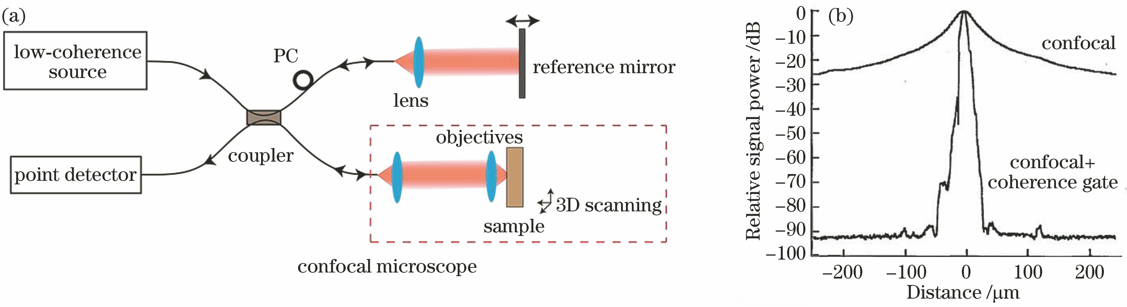
图 2. FD-OCM系统示意图及其灵敏度与TD-OCM的对比。(a) FD-OCM系统示意图;(b) 1300 nm波段高斯光源下,FD-OCM和TD-OCM灵敏度的对比[27]
Fig. 2. Schematic of FD-OCM system and comparison of sensitivity between TD-OCM and FD-OCM. (a) Schematic of FD-OCM system; (b) comparison of sensitivity between TD-OCM and FD-OCM with Gaussian source at 1300 nm[27]

图 5. PS-OCM和FF-OCM扫描成像方式示意图。(a) PS-OCM扫描成像方式;(b) FF-OCM扫描成像方式
Fig. 5. Illustrations of scanning imaging modes of PS-OCM and FF-OCM. (a) Scanning imaging mode of PS-OCM; (b) scanning imaging mode of FF-OCM

图 6. OCM分辨率随光源中心波长、带宽和数值孔径的变化规律的理论模拟。(a)轴向分辨率随光源中心波长和带宽变化的规律;(b)横向分辨率随光源中心波长和物镜数值孔径变化的规律
Fig. 6. Simulation of resolution varying with source central wavelength, bandwidth, and numerical aperture of objective in OCM. (a) Relation among axial resolution, source central wavelength, and bandwidth; (b) relation among lateral resolution, source central wavelength, and numerical aperture
图 7. 工业砂纸超高分辨成像图[38]。(a) 颗粒直径约为125 μm的工业砂纸;(b)普通系统的砂纸纵截面图像; (c)超高分辨系统的砂纸纵截面图像
Fig. 7. Ultrahigh resolution images of industrial sandpaper[38]. (a) Industrial sandpaper with particle size about 125 μm; (b) sandpaper longitudinal section image of general system; (c) sandpaper cross section image of ultra-high resolution system
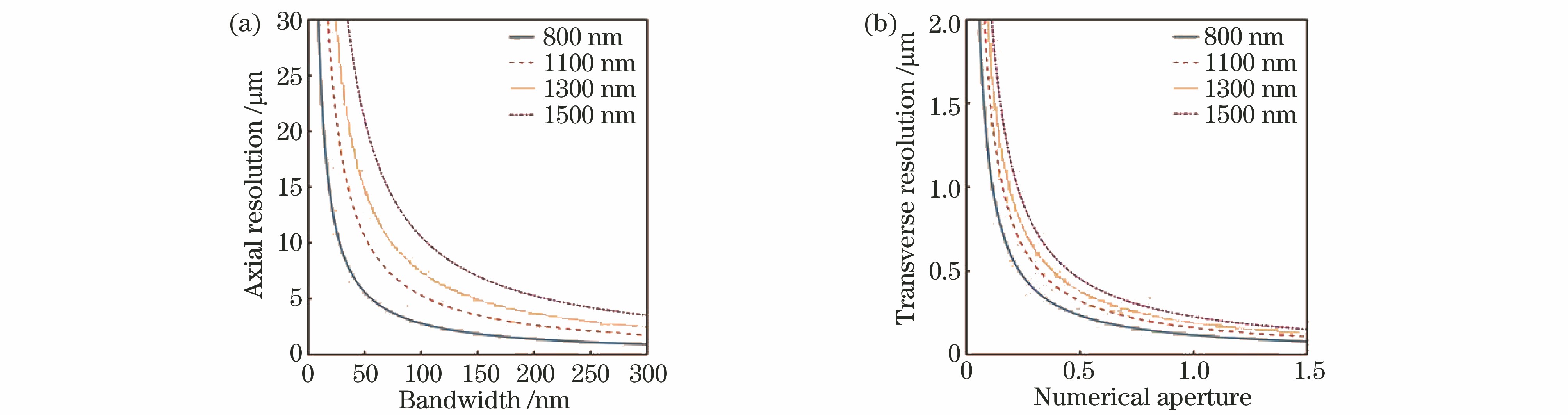
图 8. FF-OCM系统对洋葱表皮细胞高分辨成像[29]
Fig. 8. High-resolution images of onion epithelium obtained by FF-OCM system[29]
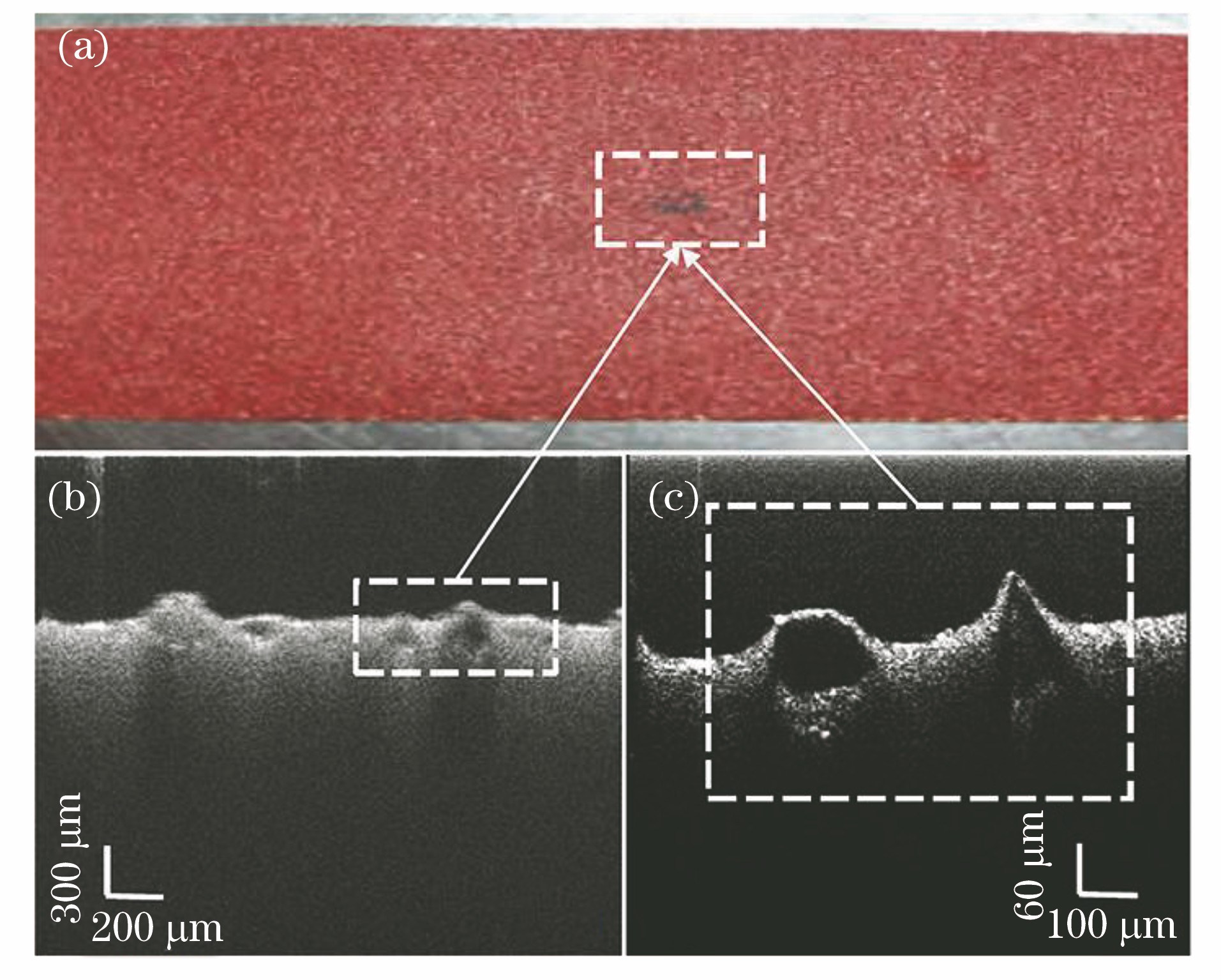
图 9. 添加随机相位前后散射样品成像图[49]。(a)未添加随机相位;(b)添加随机相位
Fig. 9. Images of scattering samples without and with random phase[49].(a) Without random phase; (b) with random phase

图 10. OCM系统轴向分辨率和探测深度、光源带宽的关系[13]。(a)在表皮组织不同深度处,轴向分辨率随高斯谱FWHM的变化;(b)表皮组织不同深度可达到的最小轴向分辨率
Fig. 10. Axial resolution of OCM system versus detection depth and source bandwidth[13]. (a) Axial resolution versus FWHM of the Gaussian spectrum at different depths of human epidermis; (b) reachable minimum axial resolution versus depth of epidermis

图 11. 暗场焦深扩展OCM样品臂示意图[13]
Fig. 11. Diagram of dark-field OCM with extended depth of focus in sample arm
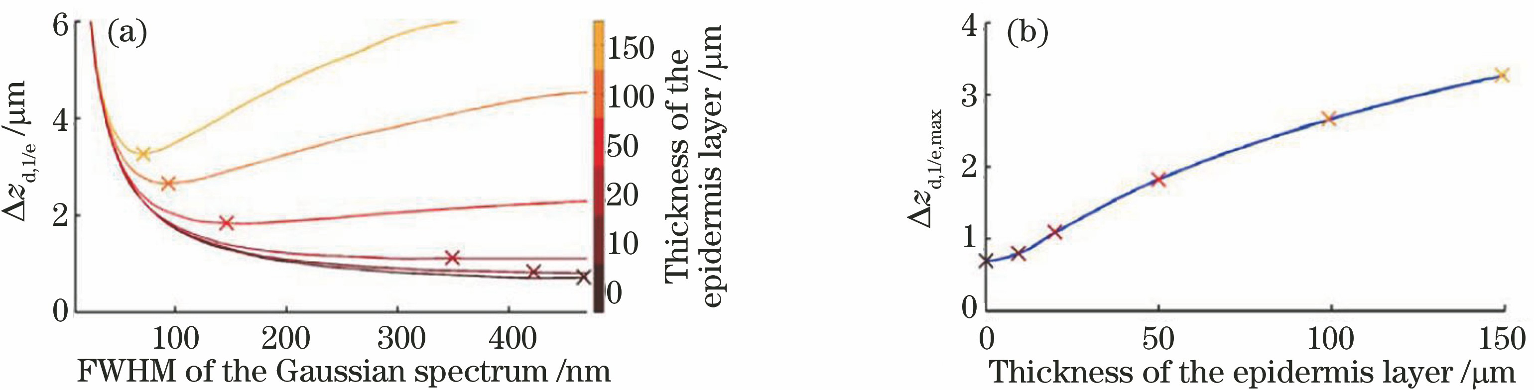
图 12. 焦深扩展OCM对小鼠胰腺组织不同深度成像的切面图[68]。比例尺:200 μm。(a) 11 μm;(b) 54 μm;(c) 97 μm;(d) 110 μm
Fig. 12. En face images of murine pancreas at different depths obtained by extended-focus OCM system[68]. Scale bar: 200 μm. (a) 11 μm; (b) 54 μm; (c) 97 μm; (d) 110 μm

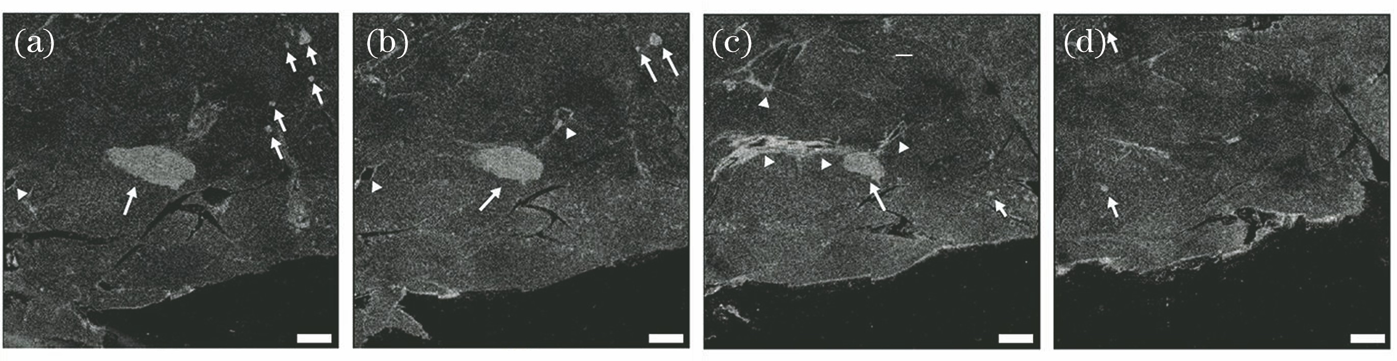
图 14. 非洲爪蛙蝌蚪截面成像[70]。(a) GD-OCM系统成像图;(b)固定焦距OCM系统成像图
Fig. 14. Cross-section images of African frog tadpole[70]. (a) Image acquired by GD-OCM system; (b) image acquired by OCM system at fixed focal plane
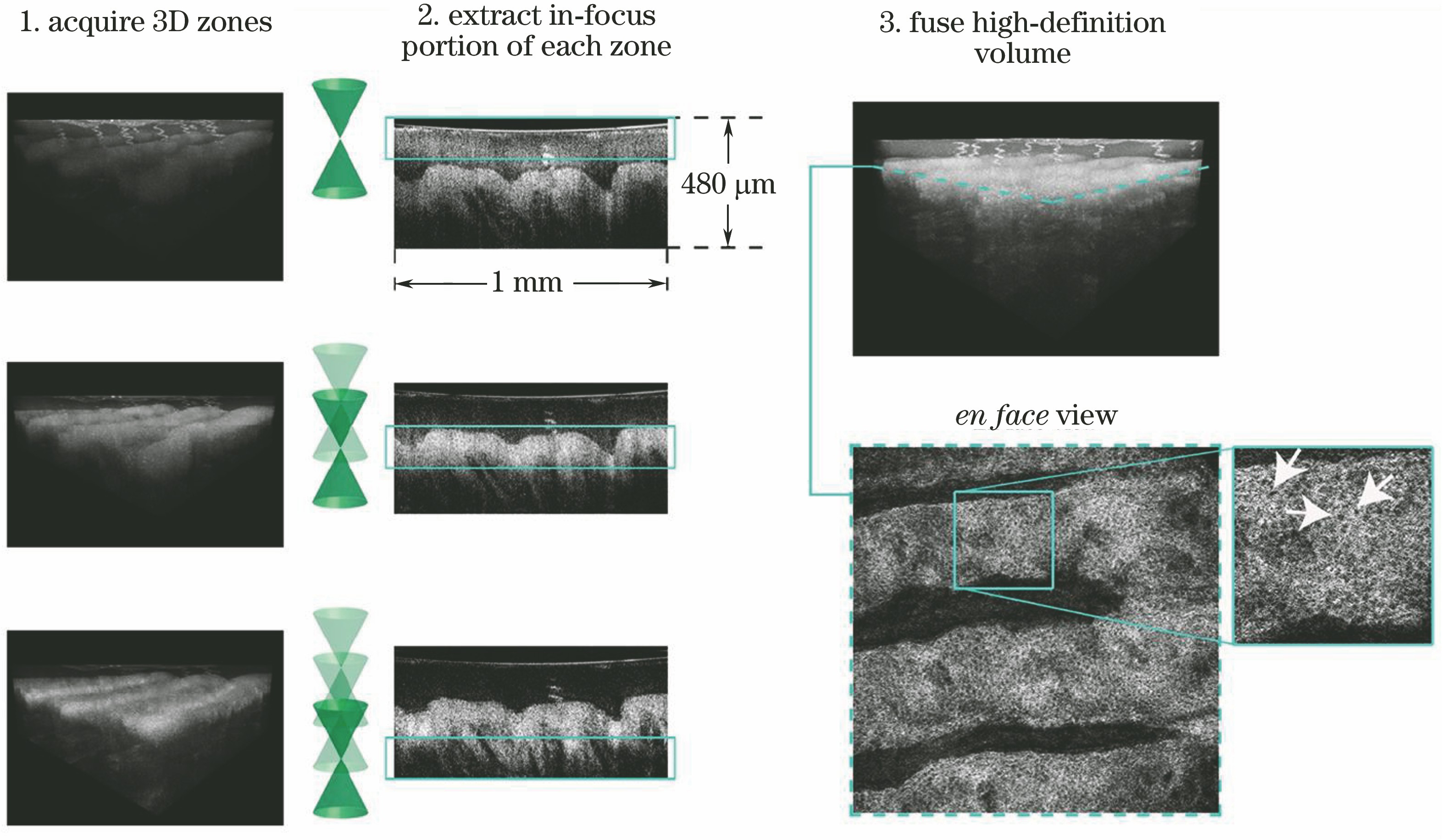
图 15. 正常角膜和FED角膜后层结构成像图[91]。(a)(c)(d)正常角膜在后弹力层、内皮细胞层、后基质层的成像;(b)(e)(f) FED角膜在后弹力层、内皮细胞层、后基质层的成像
Fig. 15. Images of posterior layers of human corneas of healthy and FED corneas[91]. (a)(c)(d) Images of healthy cornea in the posterior elastic layer, endothelial cell layer, and posterior stromal layer; (b)(e)(f) images of FED cornea in the posterior elastic layer, endothelial cell layer, and posterior stromal layer
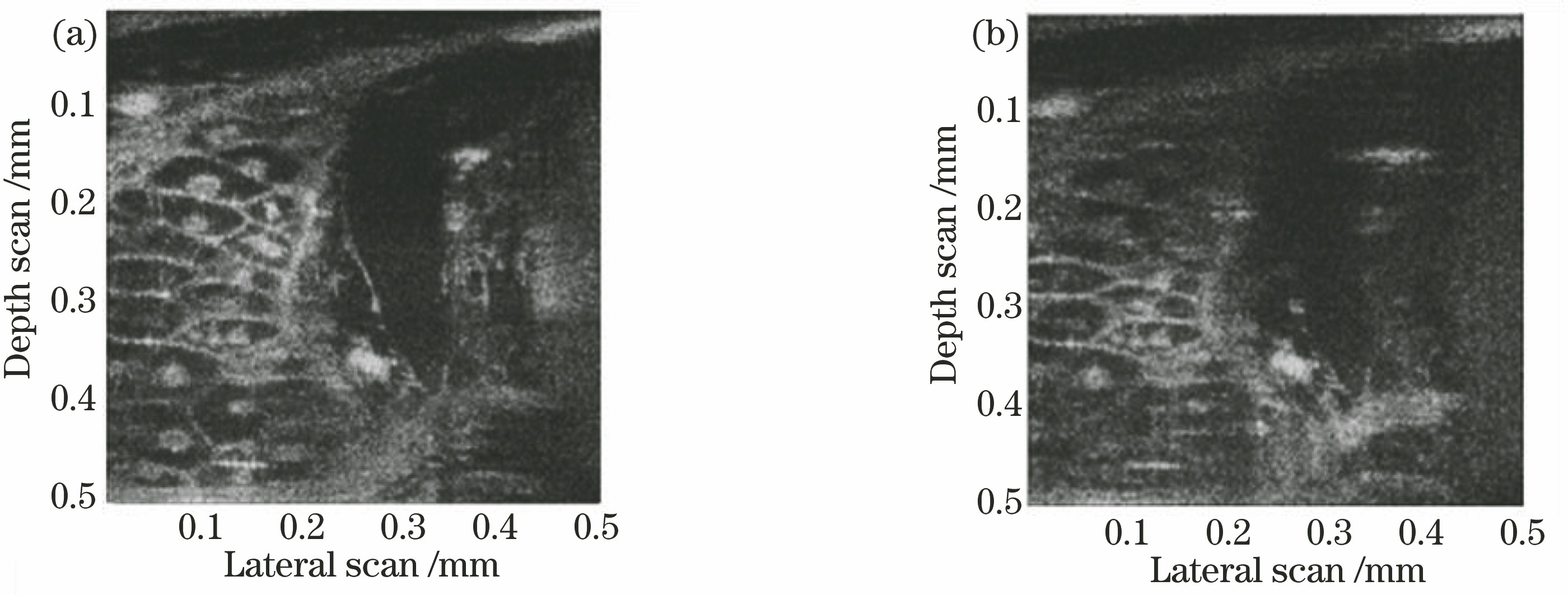
图 16. 离体小鼠脑部切片皮层组织和皮下组织成像[39]。(a)显示成像区域的透射图;(b) Vis-OCM成像结果;(c)(d)皮层组织和皮下组织的Vis-OCM成像、标记淀粉样白斑的荧光成像和叠加图
Fig. 16. Ex vivo images of cortical and subcortical structures in mouse brain slice[39]. (a) Transmission image showing imaging area; (b) result of Vis-OCM imaging; (c)(d) Vis-OCM images, fluorescence images of labeled amyloid plaques, and overlays of cortical and subcortical structures

图 17. 对纤维组织母细胞进行动态探测得到的切面成像图[110]。比例尺:50 μm。(a)切面成像图;(b)纤维伸展探测;(c)细胞迁移探测
Fig. 17. En face images of fibroblast obtained by dynamic detection[110]. Scale bar: 50 μm.(a) En face image; (b) fiber stretch detection; (c) cell migration detection
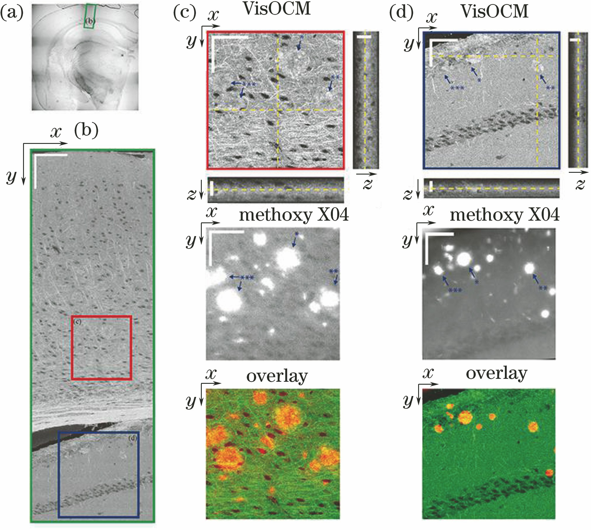
图 18. 焦深扩展OCM系统简图及其对小鼠脑皮层的血流造影图[109]。(a)焦深扩展OCM系统简图;(b)(c)血流造影图像插值处理;(d)血流造影成像图;(e)(f)总体血流流速和轴向血流流速的定量分布
Fig. 18. Diagram of focus-extended OCM and angiograms of mouse cortex[109]. (a) Illustration of focus-extended system; (b)(c) interpolation process for angiogram; (d) angiogram of mouse cortex; (e)(f) quantitative distributions of total flow rate and axial flow rate
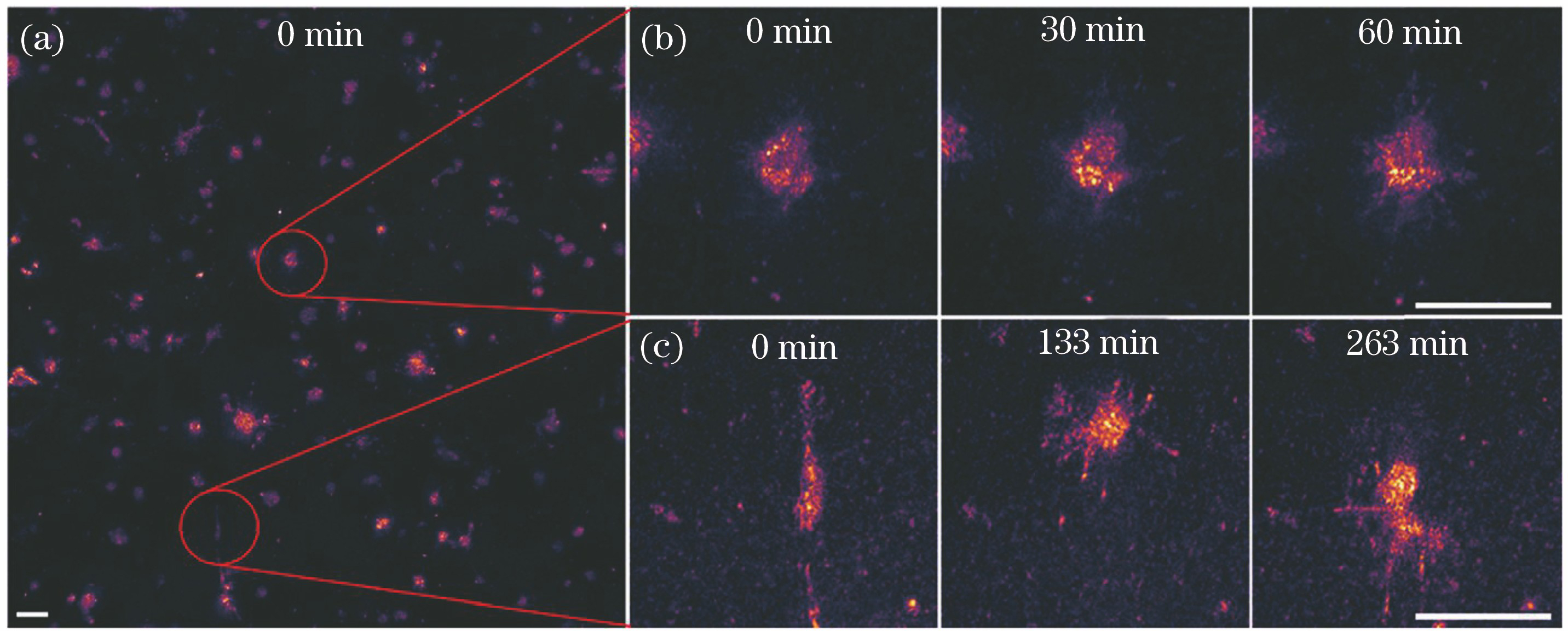
表 1各种典型成像系统的性能参数[88-90]
Table1. Typical performance parameters of different imaging systems[88-90]
|
韩涛, 邱建榕, 王迪, 孟佳, 刘智毅, 丁志华. 光学相干层析显微成像的技术与应用[J]. 中国激光, 2020, 47(2): 0207004. Han Tao, Qiu Jianrong, Wang Di, Meng Jia, Liu Zhiyi, Ding Zhihua. Optical Coherence Microscopy and Its Application[J]. Chinese Journal of Lasers, 2020, 47(2): 0207004.


 PDF全文
PDF全文

