医用不锈钢激光合金化铜钴合金的组织及其生物医学性能  下载: 939次
Microstructure and Biomedical Properties of Laser Alloyed Cu-Co Alloys on Medical Stainless Steel
图 & 表
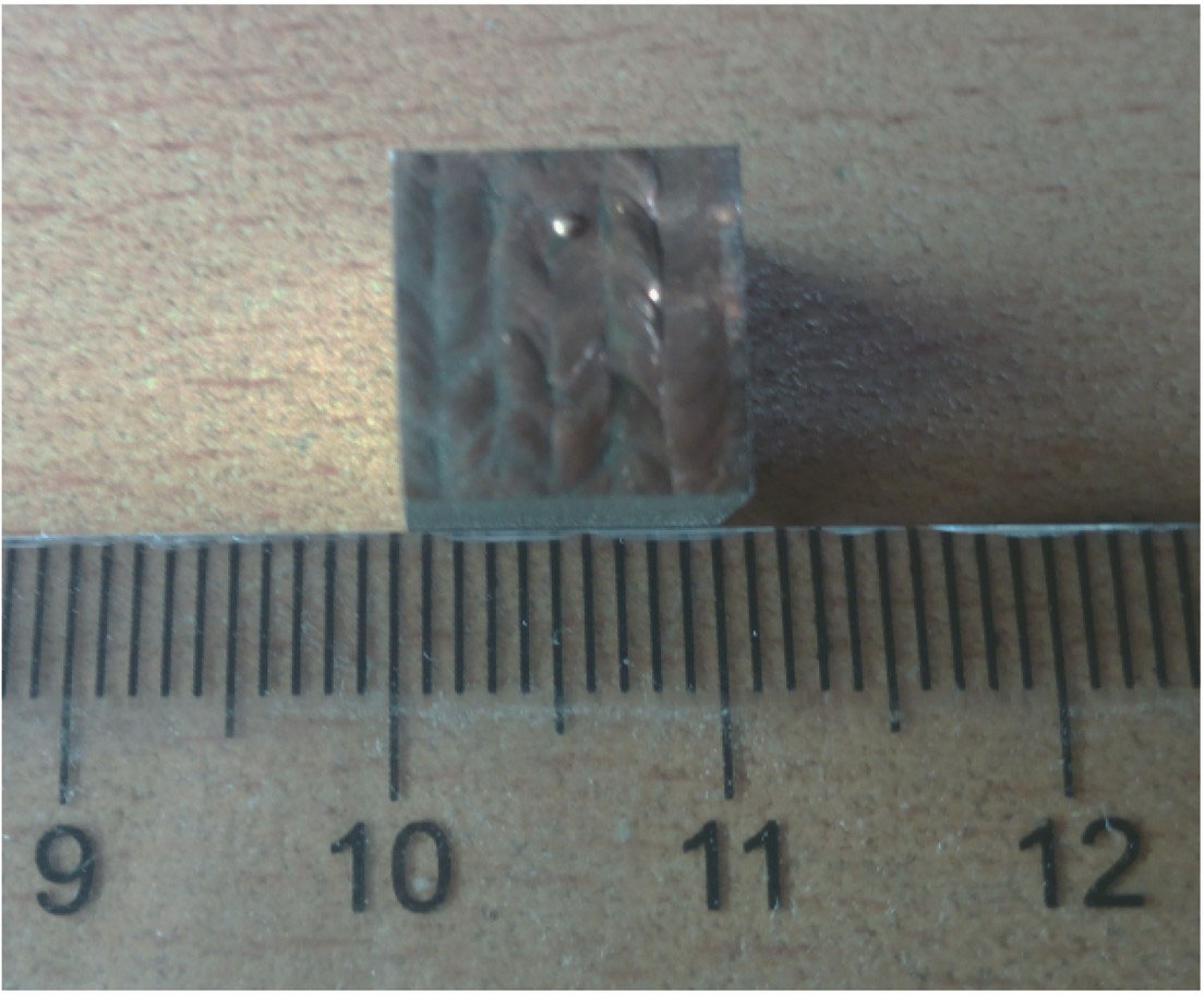
图 1. 激光合金化处理后的合金表面
Fig. 1. Alloy surface after laser alloying treatment
下载图片 查看原文
图 2. 三种比例合金的SEM形貌。(a1)样品1的低倍形貌;(a2)样品1的高倍形貌;(b1)样品2的低倍形貌;(b2)样品2的高倍形貌;(c1)样品3的低倍形貌;(c2)样品3的高倍形貌
Fig. 2. SEM morphologies of alloys with three ratios. (a1) Morphology of sample 1 at low magnification; (a2) morphology of sample 1 at high magnification; (b1) morphology of sample 2 at low magnification; (b2) morphology of sample 2 at high magnification; (c1) morphology of sample 3 at low magnification; (c2) morphology of sample 3 at high magnification
下载图片 查看原文
图 3. 三种比例合金的SEM成分扫描图样
Fig. 3. SEM scan patterns of alloys with three ratios
下载图片 查看原文
图 4. 三种金属的XRD图样以及各自峰值对应的金属成分
Fig. 4. XRD patterns of three kinds of metals and metal composition corresponding to each peak
下载图片 查看原文
图 5. 三个样品合金层中的硬度分布曲线
Fig. 5. Hardness distribution curves in alloyed layers of three samples
下载图片 查看原文
图 6. (a)无较大菌体群落的金属表面区域;(b)含较多失活的大肠杆菌群体的熔池
Fig. 6. (a) Metal surface area without large bacterial community; (b) molten pool with much dead E. coli community
下载图片 查看原文
图 7. 合金样品在腐蚀前和经3个月盐水腐蚀后通过SEM观察得到的表面形貌。(a1)腐蚀前样品1的表面形貌;(a2)腐蚀后样品1的表面形貌;(b1)腐蚀前样品2的表面形貌;(b2)腐蚀后样品2的表面形貌;(c1)腐蚀前样品3的表面形貌;(c2)腐蚀后样品3的表面形貌
Fig. 7. Surface morphologies of alloy samples observed by SEM before and after corrosion with saline water for three months. (a1) Surface morphology of sample 1 before corrosion; (a2) surface morphology of sample 1 after corrosion; (b1) surface morphology of sample 2 before corrosion; (b2) surface morphology of sample 2 after corrosion; (c1) surface morphology of sample 3 before corrosion; (c2) surface morphology of sample 3 after corrosion
下载图片 查看原文
表 1样品工艺参数
Table1. Process parameters for samples
| Sample No. | Mass ratio ofCu to Co | Presetthickness /μm | Spotdiameter /mm | Lap rate /% | Scanning speed /(mm·s-1) | Laserpower /W |
|---|
| 1 | 1∶0 | 1000 | 2.4 | 35 | 6 | 600 | | 2 | 1∶1 | 500 | 2.4 | 35 | 4 | 600 | | 3 | 2∶1 | 400 | 2.4 | 35 | 5 | 600 |
|
查看原文
表 2SEM成分扫描得到各个成分的准确占比
Table2. Accurate proportion of each component by SEM component scan
| SampleNo. | Proportionof C /% | Proportionof Mn /% | Proportionof Fe /% | Proportionof Co /% | Proportionof Cu /% |
|---|
| 1 | 2.01 | 6.22 | 68.68 | 5.29 | 17.79 | | 2 | 3.93 | 7.76 | 76.76 | 8.23 | 3.33 | | 3 | 4.67 | 5.47 | 71.05 | 10.40 | 8.41 |
|
查看原文
表 3XRD各样品峰值以及所在角度
Table3. Peak value and corresponding angle of each sample by XRD
| SampleNo. | Peak 1 | Peak 2 | Peak 3 | Peak 4 | Peak 5 |
|---|
| Angle /(°) | Value | Angle /(°) | Value | Angle /(°) | Value | Angle /(°) | Value | Angle /(°) | Value |
|---|
| 1 | 43.3 | 4048.3 | 50.4 | 1266.1 | 74.1 | 530.6 | 89.7 | 359.8 | 95.0 | 116.8 | | 2 | 43.6 | 2587.6 | 50.6 | 511.2 | 74.6 | 289.6 | 90.4 | 188.1 | 95.9 | 125.9 | | 3 | 43.5 | 2952.4 | 50.5 | 443.1 | 74.3 | 363.5 | 90.1 | 206.8 | 95.5 | 139.5 |
|
查看原文
表 4金属离子析出实验参数
Table4. Experimental parameters for metal ion precipitation
| Sample No. | Soak time /d | Concentrationfactor | Volume afterconcentration /mL | Volume ofNaOH /mL | Precipitation |
|---|
| 1 | 28 | 50 | 9 | 0.5 | Without | | 2 | 28 | 50 | 11 | 0.5 | Without | | 3 | 28 | 50 | 12 | 0.5 | Without |
|
查看原文
孙桂芳, 陶丰, 姜波, 姬文宣, 牛牧遥, 沈旭婷. 医用不锈钢激光合金化铜钴合金的组织及其生物医学性能[J]. 中国激光, 2018, 45(12): 1202008. Guifang Sun, Feng Tao, Bo Jiang, Wenxuan Ji, Muyao Niu, Xuting Shen. Microstructure and Biomedical Properties of Laser Alloyed Cu-Co Alloys on Medical Stainless Steel[J]. Chinese Journal of Lasers, 2018, 45(12): 1202008.


 PDF全文
PDF全文

