中国激光, 2018, 45 (2): 0207010, 网络出版: 2018-02-28
双光子荧光寿命成像在肿瘤诊断研究中的应用  下载: 2378次特邀综述
Applications of Two-Photon Excitation Fluorescence Lifetime Imaging in Tumor Diagnosis
图 & 表
图 1. 常用荧光寿命检测方法的基本原理[11,13-14,16,22]。(a)频域检测技术;(b)时间相关单光子计数技术
Fig. 1. Principle of common fluorescence lifetime detection methods[11,13-14,16,22]. (a) Frequency-domain detection technique; (b) time-correlated single photon counting (TCSPC) technique
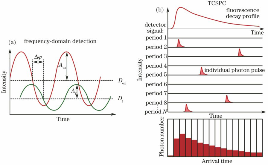
图 2. 典型的双光子荧光寿命成像系统示意图
Fig. 2. Schematic of typical two-photon excitation fluorescence lifetime imaging system

图 3. 基于仓鼠颊囊口腔癌模型活体获取的诊断为(a)正常、(b)低级别癌前病变和(c)高级别癌前病变组织的NADH荧光寿命编码图像[33];基于NADH信号获取的(d)正常的和(e)癌变的人结肠隐窝组织的离体成像结果;基于FAD信号获取的(f)正常的和(g)癌变的人结肠隐窝组织的离体成像结果[45]
Fig. 3. NADH lifetime-coded images of tissues diagnosed as (a) normal, (b) low-grade precancer, and (c) high-grade precancer acquired from the hamster cheek pouch model of oral cancer in vivo[33]; ex vivo imaging of healthy (d) and cancerous (e) human colonic crypts based on NADH; ex vivo imaging of (f) healthy and (g) cancerous human colonic crypts based on FAD[45]

图 4. 正常和癌变人体胃黏膜组织的(a~c)荧光寿命编码图像和(d~i)荧光光谱编码图像。(a)、(d)、(g)正常组织;(b)、(e)、(h)肠型腺癌;(c)、(f)、(i)神经内分泌癌
Fig. 4. (a-c) Fluorescence lifetime-coded images and (d-i) fluorescence spectra-coded images of normal and cancerous human gastric mucosa. (a), (d), (g) normal tissue; (b), (e), (h) intestinal-type adenocarcinoma; (c), (f), (i) neuroendocrine carcinoma

图 5. (a~c)小鼠神经胶质瘤边界离体成像和(d~g)人胶质母细胞瘤术中成像。(a)荧光强度图;(b)荧光寿命双色编码图;(c)正常区域和肿瘤区域荧光寿命概率分布曲线[34];(d)蛛网膜荧光强度图;(e)蛛网膜荧光寿命编码图;(f)胶质母细胞瘤的荧光强度图;(g)胶质母细胞瘤的荧光寿命编码图
Fig. 5. (a-c) Ex vivo imaging of the tumor-to-brain interface of mouse glioma and (d-g) intraoperative imaging of a human glioblastoma. (a) Fluorescence intensity image; (b) two color-coded fluorescence lifetime image; (c) Fluorescence lifetime probability distribution histograms; (d) fluorescence intensity image of arachnoid; (e) fluorescence lifetime-coded image of arachnoid; (f) fluorescence intensity image of solid tumor; (g) fluorescence lifetime-coded image of solid tumor

图 6. (a~c)小鼠正常脑组织、(d~f)小鼠神经胶质瘤皮下肿瘤组织的离体成像结果以及(g~k)小鼠原位神经胶质瘤与其周边正常脑组织的活体成像及分析结果。(a)(d)荧光强度图;(b)(e)荧光寿命编码图;(c)(f)荧光光谱编码图;(g)肿瘤周边正常脑组织的荧光强度图;(h)肿瘤边界的荧光强度图;(i)肿瘤的荧光强度图;(j) NADH荧光寿命;(k)荧光光谱
Fig. 6. Ex vivo imaging of(a-c) normal brain tissue and (d-f) subcutaneous glioma tissue and (g-k) in vivo imaging of orthotopic glioma and glioma-adjacent brain. (a)(d) Fluorescence intensity images; (b)(e) fluorescence lifetime-coded images; (c)(f) fluorescence spectra-coded images; (g) fluorescence intensity image of tumor-adjacent brain tissue; (h) fluorescence intensity image of tumor-to-brain interface; (i) fluorescence intensity image of tumor; (j) NADH fluorescence lifetime; (k) fluorescence spe

图 7. (a~d)处于不同发展阶段的小鼠黑色素瘤组织的荧光寿命编码图像以及(e~h)人离体基底细胞癌样本不同深度的荧光寿命编码图像(间隔深度为30 μm)[55]。(a)正常组织;(b) <0.5 mm的肿瘤(约12 d);(c) 1.5 mm肿瘤(约20 d);(d) 2.0~2.5 mm肿瘤(约22 d)[54]
Fig. 7. (a-d) Fluorescence lifetime-coded images of freshly excised mouse ear skin for different stages of melanoma development and (e-h) fluorescence lifetime-coded images of excised human sample of basal cell carcinoma with different depth (step size of 30 μm)[55]. (a) Normal tissue; (b) <0.5 mm lesion (about 12 days); (c) 1.5 mm lesion (about 20 days); (d) 2.0-2.5 mm lesion (about 22 days)[54]

李慧, 夏先园, 陈廷爱, 余佳, 李曦, 郑炜. 双光子荧光寿命成像在肿瘤诊断研究中的应用[J]. 中国激光, 2018, 45(2): 0207010. Li Hui, Xia Xianyuan, Chen Tingai, Yu Jia, Li Xi, Zheng Wei. Applications of Two-Photon Excitation Fluorescence Lifetime Imaging in Tumor Diagnosis[J]. Chinese Journal of Lasers, 2018, 45(2): 0207010.


 PDF全文
PDF全文

