光学学报, 2018, 38 (2): 0215003, 网络出版: 2018-08-30
基于贝叶斯理论的手臂静脉线跟踪方法  下载: 786次
Line Tracking Method of Arm Vein Based on Bayesian Theory
图 & 表
图 1. 初始种子点的确定。(a)近红外图像;(b) Gabor滤波后的图像;(c)栅格线及候选初始种子点;(d)有效的初始种子点
Fig. 1. Determination of initial seed points. (a) NIR image; (b) image after Gabor filtering; (c) grid lines and candidate initial seed points; (d) effective initial seed points
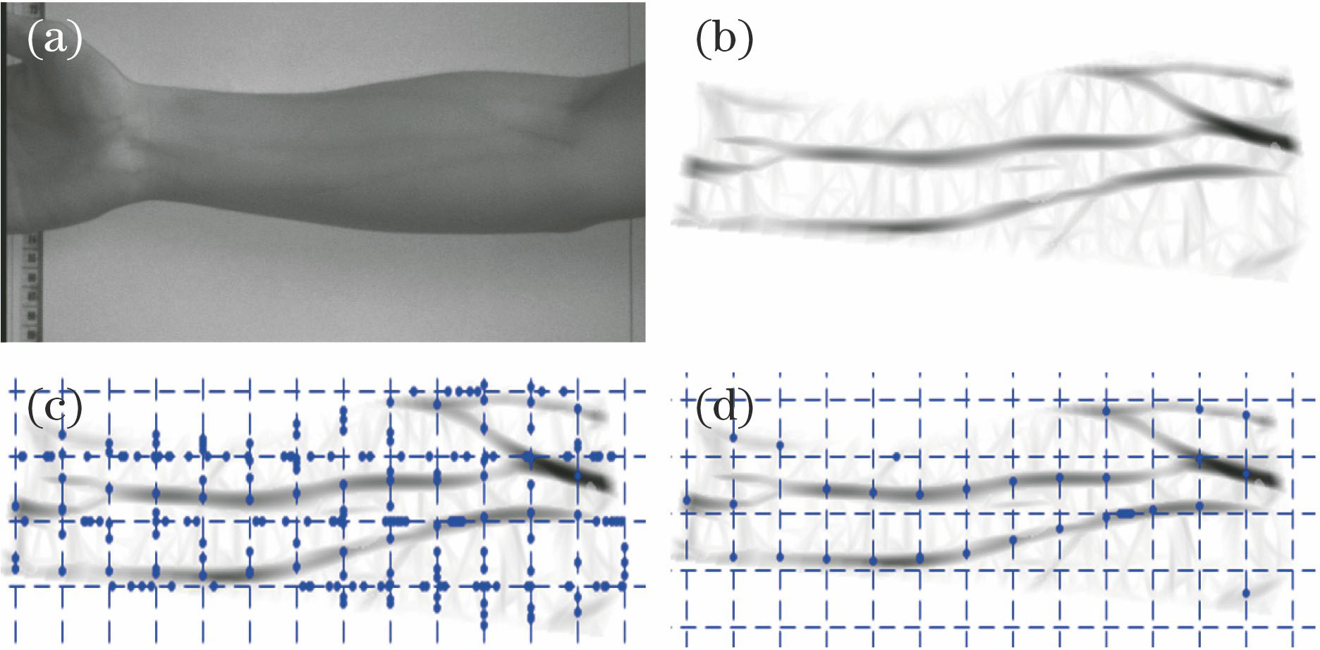

图 3. 部分初始种子点及其对应的局部血管方向
Fig. 3. Partial initial seed points and their corresponding local vessel directions

图 4. 3种类型的血管结构。(a)正常型;(b)分支型;(c)交叉型
Fig. 4. Three types of vessel structures. (a) Normal; (b) bifurcation; (c) crossing


图 7. 跟踪结果。(a)近红外图像;(b) Gabor滤波后图像;(c)跟踪到的血管边界点;(d)正常型血管边界点;(e)分支型血管边界点;(f)交叉型血管边界点
Fig. 7. Tracking results. (a) NIR image; (b) image processed with Gabor filter; (c) tracked vessel edge points; (d) edge points of normal vessel; (e) edge points of bifurcation vessel; (f) edge points of crossing vessel

图 8. 3种方法的结果对比。(a) NIR图像1;(b) NIR图像2;(c)经Gabor滤波器处理后的图像1;(d)经Gabor滤波器处理后的图像2;(e)经所提算法处理后的图像1;(f)经所提算法处理后的图像2;(g)经RLT处理后的图像1;(h)经RLT处理后的图像2;(i)经LAT处理后的图像1;(j)经LAT处理后的图像2
Fig. 8. Comparison of results of three methods. (a) NIR image 1; (b) NIR image 2; (c) image 1 processed with Gabor filter; (d) image 2 processed with Gabor filter; (e) image 1 processed with proposed method; (f) image 2 processed with proposed method; (g) image 1 processed with RLT; (h) image 2 processed with RLT; (i) image 1 processed with LAT; (j) image 2 processed with LAT

图 9. 3种方法提取结果的ROC图像。(a)所提算法;(b)重复线跟踪法;(c)局部自适应阈值法
Fig. 9. ROC images extracted by three methods. (a) Proposed method; (b) RLT method; (c) LAT method


表 13种方法的检测率对比
Table1. Comparison of detection rate of three methods
|
高昊昇, 唐超颖, 陈晓腾, 余笑. 基于贝叶斯理论的手臂静脉线跟踪方法[J]. 光学学报, 2018, 38(2): 0215003. Haosheng Gao, Chaoying Tang, Xiaoteng Chen, Xiao Yu. Line Tracking Method of Arm Vein Based on Bayesian Theory[J]. Acta Optica Sinica, 2018, 38(2): 0215003.


 PDF全文
PDF全文

