Chinese Optics Letters, 2017, 15 (5): 051701, Published Online: Jul. 23, 2018
Experimental assessment of a 3-D plenoptic endoscopic imaging system  Download: 1328次
Figures & Tables
Fig. 1. Schematic of the endoscopic setup with the plenoptic camera and relay lens system.
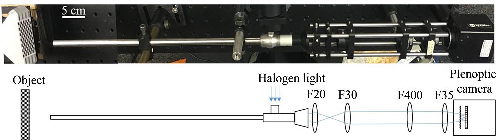


Fig. 4. (a) Microlens image of the checker board at 0° with (b) its depth map and (c, d) point cloud data at different views.

Fig. 5. Microlens image of the checker board at 45° with (b) its depth map and (c, d) point cloud data at different views.

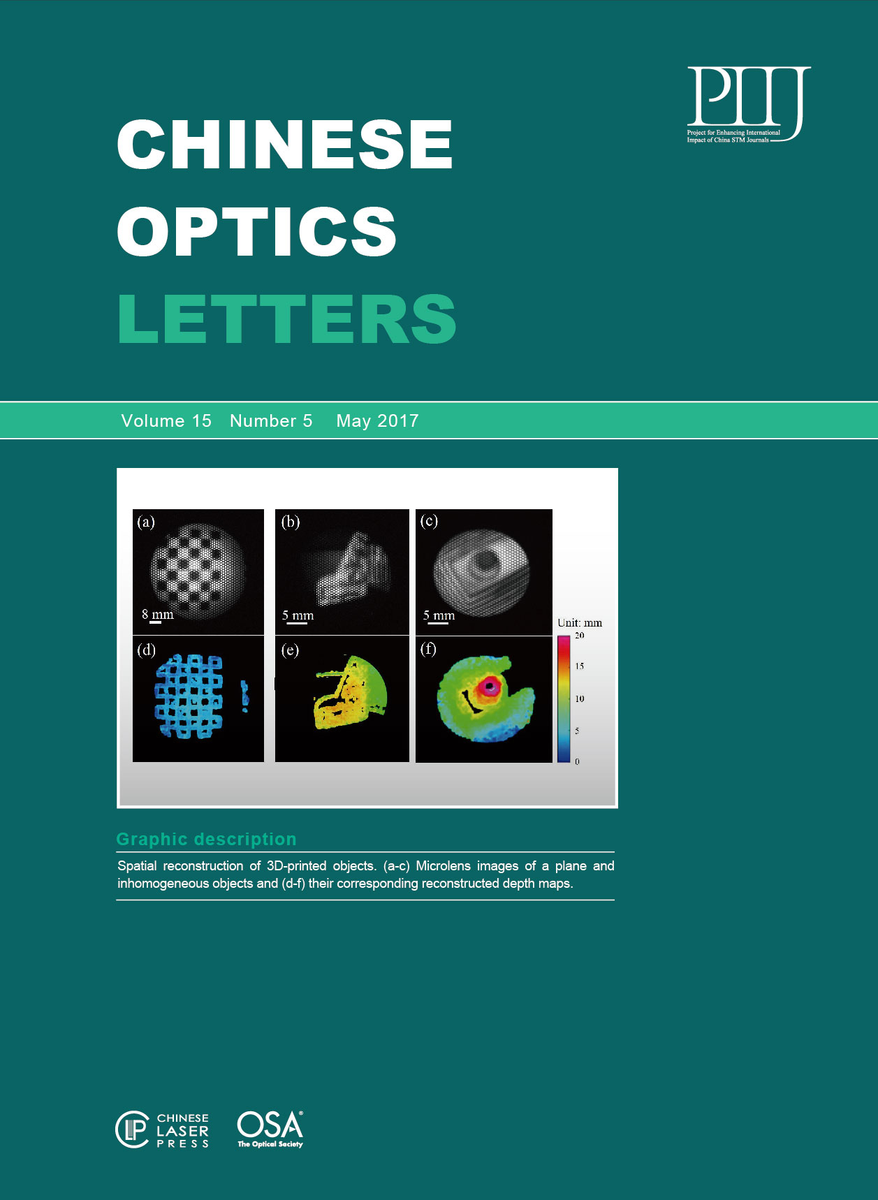
Fig. 6. (a–c) Microlens image of a plane and inhomogeneous objects and (d–f) its reconstructed depth maps.
Table1. Reconstruction Accuracy and Precision at Two Planar Angle Deviations at 0° and 45° (Unit: mm)
|
Hanh N. D. Le, Ryan Decker, Axel Krieger, Jin U. Kang. Experimental assessment of a 3-D plenoptic endoscopic imaging system[J]. Chinese Optics Letters, 2017, 15(5): 051701.


 PDF全文
PDF全文

