中国激光, 2018, 45 (5): 0510006, 网络出版: 2018-05-02   

基于空气悬浮芯微结构光纤的高灵敏度荧光检测系统 下载: 791次

High Sensitivity Fluorescence Detection System Based on Air Suspended Core Microstructural Fiber
作者单位
北京工业大学激光工程研究院, 北京市激光应用技术工程技术研究中心, 北京 100124
摘要
当空心微结构光纤纤芯的尺寸和波长相近时, 光在纤芯中的传输大大增强, 并会在纤芯周围产生很强的倏逝场。报道了一种新型的纤芯直径仅为2 μm的空气悬浮芯微结构光纤, 该光纤通过薄片堆积法拉制而成, 具有大倏逝场和微米级孔径的单元结构, 在532 nm波长处的损耗为0.16 dB/cm, 非常适合用于生化物质的传感探测。以该光纤作为传感探针, 结合激光技术搭建了一套简易的荧光光谱探测系统, 使用此系统对纳升量级的生物荧光标记材料CdTe/CdS/ZnS量子点进行荧光探测分析。利用该系统可探测荧光量子点的极限约为1 nmol/L, 相当于3.78×10⁷个量子点, 实现了高灵敏度、快速探测。基于空气悬浮芯微结构光纤的荧光检测系统为量子点标记的生物材料的灵敏检测提供了新的方法和思路。
Abstract
When the core size of a holey microstructural fiber is close to the wavelength, the light propagation in the core is greatly enhanced and a strong evanescent field is produced around the core. A new air suspended core microstructural fiber with the core diameter of only 2 μm is reported. The air suspended core fiber fabricated by sheet-stacking method has large evanescent field and the unit structure with micrometer-size holes. The air suspended core fiber, whose loss is 0.16 dB/cm at the wavelength of 532 nm, is very suitable for sensing detection of biochemical substances. Using the air suspended core fiber as sensing probe, we establish a simple fluorescence detection system combining with laser technology, and detect the fluorescence properties of bioluminescent labeling material CdTe/CdS/ZnS quantum dots with this system. The limit of the fluorescence quantum dots detected by this system is about 1 nmol/L (corresponds to 3.78×10⁷ quantum dots), which indicates that this system has the properties of high sensitivity and rapid detection speed. The fluorescence detection system based on the air suspended core microstructural fiber provides a new method and idea for the sensitive detection of biomaterials marked by quantum dots.

1 引言

目前,光纤传感技术在铁路、桥梁、石油、生物化学[1-2]等行业有着非常广泛的应用。随着光纤制造技术的发展,微结构光纤因其优异的特性而在生物化学传感探测方面备受瞩目。基于倏逝场技术的微结构光纤可以同时实现长距离信号传输和高灵敏度探测。微结构光纤具有微米级别的微孔,这种微孔可作为待测物的微型储存室,从而使待测物避免外界环境的干扰[3]。另外,微结构光纤可以由单种材料拉制而成,这相较于普通光纤可以避免外界温度和化学物质的干扰[4]

近几年,通过改进微结构光纤的结构与材料,使光场与待测物的重合得到了大幅增加,与此同时也提升了微结构光纤的检测灵敏度。例如,光子带隙型光纤(PBGF)[5-13]的特殊结构使其倏逝场与微孔中待测物的相互作用重合度得到了大幅提升,从而增加了其在传感探测方面的灵敏度。相较于拉锥纳米线光纤[14-19],PBGF具有相互作用距离长、待测物需求量少、易操作、避免环境干扰等优势。但是由于大多数PBGF存在相对有限的传输带宽,所以探测时需要激发波长与发射波长比较接近,这大大限制了此类光纤的应用范围。在此情况下,更多特种微结构光纤涌现出来。例如:英国南安普顿大学发明了一种纤芯大小为光波长的悬浮芯特种微结构光纤,此光纤具有较宽的传输波段,在生物化学传感方面具有非常广阔的应用前景[20-21];澳大利亚阿德莱德大学的Monro研究组研制了芯径在微米结构的多孔微结构光纤,并利用该光纤进行了一系列生物、化学、材料方面的高灵敏度探测[22-27]

本文介绍了一种新型的微结构光纤——空气悬浮芯微结构光纤,此光纤的纤芯直径约为2 μm。因为纤芯尺寸和光波长接近,当光在纤芯中传输时,纤芯周围就会产生较强的倏逝场。通过检测倏逝场与待测物相互作用产生的信号可以实现对待测物的检测。本课题组以这种新型微结构光纤作为传感探针搭建了一套荧光光谱探测仪,并利用此探测仪成功实现了对量子点荧光光谱的传感探测。因为量子点是一种重要的生物标记材料[24-25],该技术将为生物分子(比如蛋白质、核酸等)的灵敏检测提供新思路。

2 光纤拉制与损耗测试

空气悬浮芯微结构光纤预制棒以硼硅酸盐软玻璃材料为主,通过薄片堆积法[20]制备而成,其中纤芯和支撑片的材料分别为Schott N-BK7和Schott D263T玻璃,如图1(a)所示,预制棒的外壁由4片2 mm厚的N-BK7玻璃板拼接而成,在外壁的两侧使用切割机加工出1 mm深、200 μm宽的小槽;然后将50 μm厚、22 mm宽的Schott D263T硼硅酸盐玻璃片镶嵌到外壁两侧的沟槽中,一根由N-BK7玻璃棒拉制而成的边长为(720±10) μm的纤芯放置在玻璃片中心。堆积好的预制棒需在600~700 ℃下保温1 h,然后在玻璃转换温度(557 ℃)下进行2 h的淬火处理,以消除预制棒的内应力。

预制棒在拉丝塔上进行预拉制得到小尺寸的预制棒,图1(b)为其扫描电镜(SEM)图,然后使用外径为10 mm、内径稍大于预制棒的Schott LLF1玻璃套管将预制棒镶嵌在里面,此结构就是空气悬浮芯光纤拉丝前的预制棒结构。

图 1. (a)采用堆积法制备的预制棒;(b)预制棒的扫描电镜(SEM)图

Fig. 1. (a) Image of preform prepared by stack method; (b) SEM image of preform

下载图片 查看所有图片

制作好的预制棒最终在光纤拉丝塔上拉制形成空气悬浮芯光纤,一次性可以拉制100 m长的光纤。悬浮芯光纤的横截面如图2所示,图2(a)、(b)为不同放大倍率下悬浮芯光纤在光学显微镜下的横截面导光图,可以看出光纤导光良好,光纤外径约为200 μm;图2(c)、(d)为不同放大倍率下横截面的SEM图,光纤纤芯为类椭圆形结构,纤芯在x方向上的宽度约为2 μm,在y方向的高度约为2 μm。纤芯被一片厚度约为500 nm的薄壁支撑,纤芯两侧是等效直径约为20 μm的微孔。

图 2. 空气悬浮芯光纤横截面的形貌。(a)(b)光学显微镜图;(c)(d) SEM图

Fig. 2. Cross-section images of air suspended core fiber. (a)(b) Optical microscopy images; (c)(d) SEM images

下载图片 查看所有图片

光纤拉制成功后,使用Cutback方法测量悬浮芯光纤在532 nm波长处的传输损耗,结果如图3所示,悬浮芯光纤在532 nm波长处的传输损耗约为0.16 dB/cm。

图 3. 悬浮芯光纤在532 nm波长处的传输损耗图

Fig. 3. Propagation loss of air suspended core fiber at the wavelength of 532 nm

下载图片 查看所有图片

通过上述分析可以得出,由于悬浮芯光纤的纤芯尺寸与光波长相当,所以当光在纤芯中传输时,纤芯周围就会产生较强的倏逝场,此倏逝场会与纤芯周围的待测物相互作用而产生检测信号。另外,悬浮芯光纤自带微米级微孔,所需待测物的体积仅为纳升量级,所以可以推知这种空气悬浮芯光纤在探测生化物质方面具有灵敏度高、所需待测物体积小(纳升量级)、可避免污染等优势。

3 实验装置

利用上述光纤作为探针搭建了一套荧光光谱探测系统,实验装置如图4所示。采用前向探测法对不同浓度的生物标记材料CdTe/CdS/ZnS核壳量子点的荧光光谱进行探测分析。该量子点的粒径范围为2~10 nm,量子产率不低于50%。实验选用532 nm连续波长(CW)固体激光器作为激发光源,激光首先经过连续衰减片进行激光功率连续可调,然后通过2个反射银镜调整激光的空间光路,空气悬浮芯光纤被固定在三维调整架上,用以精确调节光纤的三维空间位置,随后激光经过焦距f为3.1 mm的非球面透镜进行空间聚焦,将激光耦合进悬浮芯光纤的纤芯中,光纤的耦合效率可以维持在40%左右。当激光在光纤纤芯中传输时,纤芯周围的倏逝场与微孔中量子点溶液相互作用产生荧光信号,在光纤的另一端使用40倍物镜将产生的信号光进行收集准直,之后再通过550 nm长通滤波片进行抽运光滤除,进而耦合进海洋光学光谱仪的多模光纤中。

图 4. 实验装置示意图

Fig. 4. Schematic of experimental setup

下载图片 查看所有图片

实验中使用的悬浮芯光纤长度约为10 cm,待测物量子点悬浮液通过毛细作用被吸入悬浮芯光纤的微孔中,待测物充满整个光纤所需时间不到30 s,计算得到光纤充满时所需液体仅为62 nL左右,悬浮芯光纤填充前后的纵截面对比如图5所示。由于折射率的改变,当悬浮芯光纤未填充量子点悬浮液时,光纤的微孔区呈黑色,如图5(a)所示;充入量子点悬浮液后,微孔区呈透明状,如图5(b)所示。

图 5. 悬浮芯光纤(a)未充入和(b)充入量子点悬浮液后的纵截面

Fig. 5. Longitudinal section of air suspended core fiber (a) unfilled and (b) filled with quantum dots suspension

下载图片 查看所有图片

4 实验结果与分析

在使用悬浮芯光纤探测量子点荧光光谱之前,首先在酶标仪上测量量子点的荧光光谱,所测量子点悬浮液的浓度为1 μmol/L,设量子点的激发波长为532 nm,测量结果如图6所示。由图6可以看出,量子点的荧光光谱范围为560~800 nm,荧光峰值波长约为665 nm,光谱的半峰全宽(FWHM)为70 nm。

接下来使用图4的装置分别对浓度分别为

图 6. 酶标仪测得的量子点的荧光光谱

Fig. 6. Fluorescence spectrum of quantum dots measured by a microplate reader

下载图片 查看所有图片

100,50,10,5,2.5 nmol/L的量子点悬浮液进行探测,光纤探针长度为(10±1) cm,激光抽运功率为340 μW左右。为了消除因光纤端面切割不均匀而导致的误差,通过调整抽运功率来保证耦合进光纤纤芯中的功率相同。在光纤长度相同的情况下,保证光纤末端的输出功率相同。本实验设定末端功率为100 μW,光谱仪探测积分时间为1 s。

实验中首先对浓度为100 nmol/L的量子点悬浮液进行探测,测得的量子点的荧光光谱如图7所示。荧光光谱范围为580~800 nm,荧光峰值波长为685 nm,与图6所示的采用酶标仪测得的峰值波长(665 nm)相比出现了22 nm左右的红移,这是荧光分子在光纤传输过程中产生重吸收效应所导致的光谱红移现象。然后分别对50,10,5,2.5 nmol/L量子点悬浮液进行探测,测得的量子点的荧光光谱如图7所示。由图7可见:随着量子点悬浮液浓度降低,荧光光谱的强度随之减弱;当量子点悬浮液浓度降低到2.5 nmol/L时,已无法分辨信号和背景噪音。图7(b)是归一化后的光谱图,可以看出,随着量子点悬浮液的浓度降低,荧光光谱有展宽的趋势,这是因为随着荧光强度的减弱,光纤的背景荧光对光谱的影响愈加明显。

图 7. 悬浮芯光纤测得的不同浓度悬浮液中量子点的(a)原始荧光光谱和(b)归一化荧光光谱

Fig. 7. (a) Raw and (b) normalized fluorescence spectra of quantum dots in suspension with different concentrations measured by air suspended core fiber

下载图片 查看所有图片

测得的量子点荧光强度(峰值强度和面积积分强度)与浓度呈现良好的线性关系,如图8所示,计算可得检测限分别为1.38 nmol/L和0.79 nmol/L。对于长度为10 cm的悬浮芯光纤,按照检测限为1 nmol/L计算,使用前向探测法可以测得的量子点个数约为3.78×107

图 8. 悬浮芯光纤测得量子点荧光(a)峰值强度与(b) 600~800 nm面积积分强度与浓度的线性关系

Fig. 8. Linear relation between the concentration and (a) fluorescence peak intensity and (b) 600-800 nm area integral intensity of quantum dots measured by air suspended core fiber

下载图片 查看所有图片

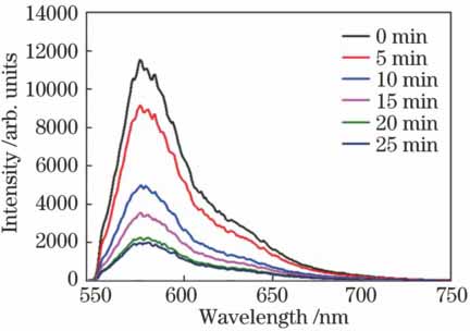
为了证明所建系统的普适性,本研究还对常用荧光染料罗丹明B的荧光光谱进行了测定,并且对罗丹明B的荧光漂白性能进行了检测,结果见图9。由图9可以看到:罗丹明B的荧光强度随时间延长呈明显下降的趋势,荧光强度衰减到50%时所用时间大约为9.5 min。

图 9. 罗丹明B的荧光漂白曲线

Fig. 9. Photo-bleaching curves of Rhodamine B

下载图片 查看所有图片

纤芯的长度代表了待测物与纤芯周围光场相互作用的纵向距离,本研究测量了不同长度悬浮芯光纤对量子点荧光强度的影响。

首先将抽运光耦合进长度为92 cm的悬浮芯光纤中,将100 nmol/L的量子点悬浮液充入光纤中,然后使用Cutback法逐次切割光纤的尾端,得到利用不同长度悬浮芯光纤测得的量子点的荧光强度,结果如图10所示。由图10可以看出:测得量子点的荧光强度随着光纤长度的减小而呈现先增加后减小的抛物线趋势,在光纤长度为50 cm左右时,荧光强度达到最大,如图10(b)所示;当光纤长度小于50 cm时,荧光强度与光纤长度呈正相关关系,这是因为随着光纤长度增加,光场与更多的待测物作用,导致测得的光谱强度不断增加;当光纤长度大于50 cm后,光纤损耗效应占主导,荧光信号减弱。

图 10. (a)不同长度光纤测得的量子点的荧光光谱;(b)荧光强度与光纤长度的拟合曲线

Fig. 10. (a) Measured fluorescence spectra of quantum dots using different lengths of fiber; (b) fitting curve of fluorescence intensity and fiber length

下载图片 查看所有图片

5 结论

首先介绍了一种新型结构的微结构光纤——空气悬浮芯光纤,然后以悬浮芯微结构光纤作为探针搭建了一套简易的荧光光谱仪,并利用这套装置采用前向探测法对生物标记材料CdTe/CdS/ZnS核壳量子点的荧光光谱进行了探测。实验结果显示,此装置对量子点的探测极限是1 nmol/L左右,即可以探测到的量子点个数约为3.78×107,这已经达到了高灵敏度探测器的水平。另外,此光纤所需的待测物体积仅为纳升量级,在探测时可使待测物与外部环境隔离,防止待测物被污染,以便进行精准探测。此装置可以用于荧光材料(量子点、荧光染料等)标记的生物分子的检测,比如病原性DNA、蛋白质或者抗体,甚至可以用于荧光标记的肿瘤细胞的检测,具有高灵敏度、所需待测物少(纳升量级)、成本低、紧凑性好等优点。

参考文献

[1] 黄惠杰, 翟俊辉, 任冰强, 等. 光纤倏逝波生物传感器及其应用[J]. 光学学报, 2003, 15(4): 652-655.

    黄惠杰, 翟俊辉, 任冰强, 等. 光纤倏逝波生物传感器及其应用[J]. 光学学报, 2003, 15(4): 652-655.

    Huang H J, Zhai J H, Ren B Q, et al. Fiber-optic evanescent wave biosensor and its applications[J]. Acta Optica Sinica, 2003, 15(4): 652-655.

    Huang H J, Zhai J H, Ren B Q, et al. Fiber-optic evanescent wave biosensor and its applications[J]. Acta Optica Sinica, 2003, 15(4): 652-655.

[2] 黄惠杰, 翟俊辉, 赵永凯, 等. 多探头光纤倏逝波生物传感器及其性能研究[J]. 中国激光, 2004, 31(6): 718-722.

    黄惠杰, 翟俊辉, 赵永凯, 等. 多探头光纤倏逝波生物传感器及其性能研究[J]. 中国激光, 2004, 31(6): 718-722.

    Huang H J, Zhai J H, Zhao Y K, et al. Multi-probe fiber-optic evanescent wave biosensor and its characterization[J]. Chinese Journal of Lasers, 2004, 31(6): 718-722.

    Huang H J, Zhai J H, Zhao Y K, et al. Multi-probe fiber-optic evanescent wave biosensor and its characterization[J]. Chinese Journal of Lasers, 2004, 31(6): 718-722.

[3] Monro T M, Belardi W, Furusawa K, et al. Sensing with microstructured optical fibres[J]. Measurement Science & Technology, 2001, 12(7): 854.

    Monro T M, Belardi W, Furusawa K, et al. Sensing with microstructured optical fibres[J]. Measurement Science & Technology, 2001, 12(7): 854.

[4] Ebendorff-Heidepriem H, Monro T M. Extrusion of complex preforms for microstructured optical fibers[J]. Optics Express, 2007, 15(23): 15086-15092.

    Ebendorff-Heidepriem H, Monro T M. Extrusion of complex preforms for microstructured optical fibers[J]. Optics Express, 2007, 15(23): 15086-15092.

[5] Jensen J B, Pedersen L H, Hoiby P E. et al. Photonic crystal fiber based evanescent-wave sensor for detection of biomolecules in aqueous solutions[J]. Optics Letters, 2004, 29(17): 760-762.

    Jensen J B, Pedersen L H, Hoiby P E. et al. Photonic crystal fiber based evanescent-wave sensor for detection of biomolecules in aqueous solutions[J]. Optics Letters, 2004, 29(17): 760-762.

[6] Smolka S, Barth M, Benson O. Highly efficient fluorescence sensing with hollow core photonic crystal fibers[J]. Optics Express, 2007, 15(20): 12783-12791.

    Smolka S, Barth M, Benson O. Highly efficient fluorescence sensing with hollow core photonic crystal fibers[J]. Optics Express, 2007, 15(20): 12783-12791.

[7] Bise RT, Windeler RS, Kranz KS, et al. Tunable photonic band gap fiber[C]. Optical Fiber Communication Conference and Exhibit, 2002: 466- 468.

    Bise RT, Windeler RS, Kranz KS, et al. Tunable photonic band gap fiber[C]. Optical Fiber Communication Conference and Exhibit, 2002: 466- 468.

[8] Hoo Y L, Jin W, Ho H L, et al. Measurement of gas diffusion coefficient using photonic crystal fiber[J]. IEEE Photonics Technology Letters, 2003, 15(10): 1434-1436.

    Hoo Y L, Jin W, Ho H L, et al. Measurement of gas diffusion coefficient using photonic crystal fiber[J]. IEEE Photonics Technology Letters, 2003, 15(10): 1434-1436.

[9] Du F, Lu Y Q, Wu S T. Electrically tunable liquid-crystal photonic crystal fiber[J]. Applied Physics Letters, 2004, 85(12): 2181-2183.

    Du F, Lu Y Q, Wu S T. Electrically tunable liquid-crystal photonic crystal fiber[J]. Applied Physics Letters, 2004, 85(12): 2181-2183.

[10] 许震宇, 张若京, 龚益玲. 光子晶体压力传感器的基本原理[J]. 物理学报, 2004, 53(3): 724-727.

    许震宇, 张若京, 龚益玲. 光子晶体压力传感器的基本原理[J]. 物理学报, 2004, 53(3): 724-727.

    Xu Z Y, Zhang R J, Gong Y L. The principles of pressure sensors based on photonic crystal[J]. Acta Physica Sinica, 2004, 53(3): 724-727.

    Xu Z Y, Zhang R J, Gong Y L. The principles of pressure sensors based on photonic crystal[J]. Acta Physica Sinica, 2004, 53(3): 724-727.

[11] MagalhaesF, Carvalho JP, Ferreira LA, et al. Methane detection system based on wavelength modulation spectroscopy and hollow-core fibres[C]. IEEE Sensors Conference, 2008: 1277- 1280.

    MagalhaesF, Carvalho JP, Ferreira LA, et al. Methane detection system based on wavelength modulation spectroscopy and hollow-core fibres[C]. IEEE Sensors Conference, 2008: 1277- 1280.

[12] Wei H, Song K, Tong W. et al. Some features of the photonic crystal fiber temperature sensor with liquid ethanol filling[J]. Optics Express, 2010, 18(15): 15383-15388.

    Wei H, Song K, Tong W. et al. Some features of the photonic crystal fiber temperature sensor with liquid ethanol filling[J]. Optics Express, 2010, 18(15): 15383-15388.

[13] 王若琪, 姚建铨, 周睿, 等. 填充混合液体的光子晶体光纤温度传感研究[J]. 光电子·激光, 2011, 22(11): 1609-1612.

    王若琪, 姚建铨, 周睿, 等. 填充混合液体的光子晶体光纤温度传感研究[J]. 光电子·激光, 2011, 22(11): 1609-1612.

    Wang R Q, Yao J Q, Zhou R, et al. Research of photonic crystal fiber temperature sensor with mixture liquid filling[J]. Journal of Optoelectronics·Laser, 2011, 22(11): 1609-1612.

    Wang R Q, Yao J Q, Zhou R, et al. Research of photonic crystal fiber temperature sensor with mixture liquid filling[J]. Journal of Optoelectronics·Laser, 2011, 22(11): 1609-1612.

[14] Lou J, Tong L, Ye Z. Modeling of silica nanowires for optical sensing[J]. Optics Express, 2005, 13(6): 2135-2140.

    Lou J, Tong L, Ye Z. Modeling of silica nanowires for optical sensing[J]. Optics Express, 2005, 13(6): 2135-2140.

[15] Polynkin P, Polynkin A, Peyghambarian N, et al. Evanescent field-based optical fiber sensing device for measuring the refractive index of liquids in microfluidic channels[J]. Optics Letters, 2005, 30(11): 1273-1275.

    Polynkin P, Polynkin A, Peyghambarian N, et al. Evanescent field-based optical fiber sensing device for measuring the refractive index of liquids in microfluidic channels[J]. Optics Letters, 2005, 30(11): 1273-1275.

[16] Chen G Y, Zhang X L, Brambilla G. et al. Theoretical and experimental demonstrations of a microfiber-based flexural disc accelerometer[J]. Optics Letters, 2011, 36(18): 3669-3671.

    Chen G Y, Zhang X L, Brambilla G. et al. Theoretical and experimental demonstrations of a microfiber-based flexural disc accelerometer[J]. Optics Letters, 2011, 36(18): 3669-3671.

[17] Villatoro J, Monzón-Hernández D. Fast detection of hydrogen with nano fiber tapers coated with ultra thin palladium layers[J]. Optics Express, 2005, 13(13): 5087-5092.

    Villatoro J, Monzón-Hernández D. Fast detection of hydrogen with nano fiber tapers coated with ultra thin palladium layers[J]. Optics Express, 2005, 13(13): 5087-5092.

[18] Zhang L, Gu F, Lou J. et al. Fast detection of humidity with a subwavelength-diameter fiber taper coated with gelatin film[J]. Optics Express, 2008, 16(17): 13349-13353.

    Zhang L, Gu F, Lou J. et al. Fast detection of humidity with a subwavelength-diameter fiber taper coated with gelatin film[J]. Optics Express, 2008, 16(17): 13349-13353.

[19] Brambilla G, Finazzi V, Richardson D J. Ultra-low-loss optical fiber nanotapers[J]. Optics Express, 2004, 12(10): 2258-2263.

    Brambilla G, Finazzi V, Richardson D J. Ultra-low-loss optical fiber nanotapers[J]. Optics Express, 2004, 12(10): 2258-2263.

[20] Shi J, Feng X, Lian Z, et al. Fabrication of multiple parallel suspended-core optical fibers by sheet-stacking[J]. Optical Fiber Technology, 2014, 20(4): 395-402.

    Shi J, Feng X, Lian Z, et al. Fabrication of multiple parallel suspended-core optical fibers by sheet-stacking[J]. Optical Fiber Technology, 2014, 20(4): 395-402.

[21] Shi J D, Feng X, Horak P. et al. A fiberized highly birefringent glass micrometer-size ridge waveguide[J]. Optical Fiber Technology, 2015, 23: 137-144.

    Shi J D, Feng X, Horak P. et al. A fiberized highly birefringent glass micrometer-size ridge waveguide[J]. Optical Fiber Technology, 2015, 23: 137-144.

[22] Ebendorff-HeidepriemH, PetropoulosP, MooreR, et al. Fabrication and optical properties of lead silicate glass holey fibers[J]. Journal of Non-Crystalline Solids, 2004, 345/346: 293- 296.

    Ebendorff-HeidepriemH, PetropoulosP, MooreR, et al. Fabrication and optical properties of lead silicate glass holey fibers[J]. Journal of Non-Crystalline Solids, 2004, 345/346: 293- 296.

[23] Afshar S, Warren-Smith S, Monro T M. Enhancement of fluorescence-based sensing using microstructured optical fibres[J]. Optics Express, 2007, 15(26): 17891-17901.

    Afshar S, Warren-Smith S, Monro T M. Enhancement of fluorescence-based sensing using microstructured optical fibres[J]. Optics Express, 2007, 15(26): 17891-17901.

[24] Ruan Y, Schartner E P, Ebendorff-Heidepriem H, et al. Detection of quantum-dot labelled proteins using soft glass microstructured optical fibers[J]. Optics Express, 2007, 15(26): 17819-17826.

    Ruan Y, Schartner E P, Ebendorff-Heidepriem H, et al. Detection of quantum-dot labelled proteins using soft glass microstructured optical fibers[J]. Optics Express, 2007, 15(26): 17819-17826.

[25] Ruan Y L, Foo T C, Warren-Smith S, et al. Antibody immobilization within glass microstructured fibers: a route to sensitive and selective biosensors[J]. Optics Express, 2008, 16(22): 18514-18523.

    Ruan Y L, Foo T C, Warren-Smith S, et al. Antibody immobilization within glass microstructured fibers: a route to sensitive and selective biosensors[J]. Optics Express, 2008, 16(22): 18514-18523.

[26] Nguyen L V, Warren-Smith S, Cooper A. et al. Molecular beacons immobilized within suspended core optical fiber for specific DNA detection[J]. Optics Express, 2012, 20(28): 29378-29385.

    Nguyen L V, Warren-Smith S, Cooper A. et al. Molecular beacons immobilized within suspended core optical fiber for specific DNA detection[J]. Optics Express, 2012, 20(28): 29378-29385.

[27] Warren-Smith S, Nie G C, Schartner E P. et al. Enzyme activity assays within microstructured optical fibers enabled by automated alignment[J]. Biomedical Optics Express, 2012, 3(12): 3304-3313.

    Warren-Smith S, Nie G C, Schartner E P. et al. Enzyme activity assays within microstructured optical fibers enabled by automated alignment[J]. Biomedical Optics Express, 2012, 3(12): 3304-3313.

张炤, 王秀翃, 乔鹏飞, 刘倩倩, Lang Marion, 冯宪, 王璞. 基于空气悬浮芯微结构光纤的高灵敏度荧光检测系统[J]. 中国激光, 2018, 45(5): 0510006. Zhang Zhao, Wang Xiuhong, Qiao Pengfei, Liu Qianqian, Lang Marion, Feng Xian, Wang Pu. High Sensitivity Fluorescence Detection System Based on Air Suspended Core Microstructural Fiber[J]. Chinese Journal of Lasers, 2018, 45(5): 0510006.
本文已被 1 篇论文引用
被引统计数据来源于中国光学期刊网
引用该论文: TXT   |   EndNote

相关论文

加载中...

关于本站 Cookie 的使用提示

中国光学期刊网使用基于 cookie 的技术来更好地为您提供各项服务,点击此处了解我们的隐私策略。 如您需继续使用本网站,请您授权我们使用本地 cookie 来保存部分信息。
全站搜索
您最值得信赖的光电行业旗舰网络服务平台!