基于内标法的表面增强拉曼散射定量分析  下载: 1769次
Quantitative Analysis of Surface-Enhanced Raman Scattering Based on Internal Standard Method
重庆大学光电工程学院光电技术及系统教育部重点实验室, 重庆 400044
图 & 表
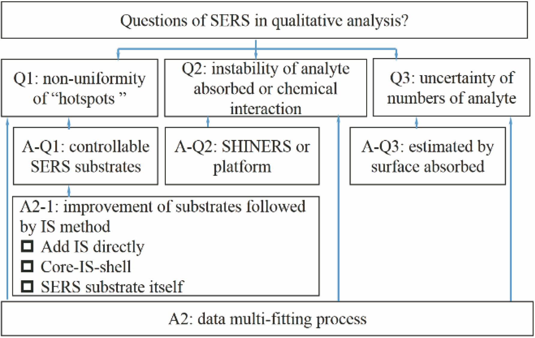
图 1. SERS定量分析中的问题
Fig. 1. Problems in SERS quantitative detection
下载图片 查看原文
图 2. 内标法示意图
Fig. 2. Schematic diagram of internal standard method
下载图片 查看原文
图 4. 壳核结构对于SRES性能的改善:(a)壳作为保护层以避免化学效应的影响[17]; (b)壳作为粒子功能化或分子吸附平台[36];(c)碳纳米材料包裹金属纳米粒子在生物科学中的应用[42-43]
Fig. 4. Shell core structure improves the performance of SERS substrate: (a)Shell acts as a protective layer to avoid the effects of chemical effects[17]; (b) shell acts as a particle functionalization or molecular adsorption platform[36]; (c) application of carbon nanomaterials wrapped metal nanoparticles in biological science[42- 下载图片 查看原文
图 5. 三种内标物添加模式。(a) A:外部添加模式,包括直接添加内标物或添加标记分子;(b) B:核-内标分子-壳模式;(c) C:自标定复合基底
Fig. 5. Three internal standard addition modes. (a) A: External addition mode, including add internal standards directly or add labeling molecules; (b) B: core-internal standard-shell modes; (c) C: self-calibrating substrate
下载图片 查看原文
表 1三种内标模式下的结构、内标物和性能
Table1. Structure, internal standard and performance in three internal standard modes
| Mode | Structure | IS | Performance (LOD or EF) | Reference |
|---|
| External addition | Silver Colloid | IEIS:isotopicR6G | 2×10⁻¹⁰ mol/L (R6G)10-9 mol/L (R6G) | Zhang et al[52]Perera et al[53] | | AuNP | ILC:Trp-d5 or13C | 10-7 mol/L(Trp and caffeine) | Subaihi et al[54] | | AuNP | TMT | 2.9×10⁻⁹ mol/L (Cd2+) | Chen et al[50] | | Microfluidics withAg nanocolloids | RhBp-thiocresol | 5×10⁻⁷ mol/L (MG)5×10⁻⁸ mol/L (MG) | Xia et al[49] | | Ag@AuNCs | NTP | EF:104, aldolcondensation reactionof acetone and MTBH | Weatherston et al[33] | | Ag films | Phosphatebackbone | adenine and cytosine | Freeman et al[8] | | Core-molecule-shell | Au@CA+Mpy@AgNPs | Mpy | 5×10⁻¹⁰ mol/L (PDI) | Shen et al[55] | | CISS NC | pMBA+pATP | 3×10-11 mol/L (R6G) | Wu et al[56] | | Au@2-MB+PATP@Ag | 2-MBPATP2-MB+PATP | 3×10⁻⁷ mol/L (Phosmet) | Zhang et al[57] | | Self-calibratingsubstrate | FONTopUp@Ag | SiSi | 0.5 mg/L (E 122)2×10⁻⁸ mol/L (SMX) | Peksa et al[61]Patze et al[32] | | AGNs | Graphitic | EF>106 (RhB) | Zou et al[59] | | G-SERS | GE | 10-8 mol/L (CV and RhB) | Tian et al[30] | | CNT/AgNPs | CNT | 10-9 mol/L (R6G) | Zhang et al[60] | | GE/AgNHs | GE | 10-15 mol/L (R6G) | Zhang et al[35] | | Note:Abbreviation:limits of detection (LOD), isotope edited internal standard (IEIS), isotopically labelled compound (ILC), enhancement factor(EF); 4-(methylthio) benzaldehyde(MTBH);Substrate:colloidal Ag-Au core-shell nanocubes (Ag@AuNCs), sulfamethoxazole (SMX), “film over nanospheres” (FON), Au@Ag nanocuboids (NCs),core-internal standard-shell (CISS);Material:Rhodamine6G (R6G), Tryptophan (Trp), malachite green (MG), 4-Nitrothiophenol (NTP), trithiocyanuric acid (TMT), 2-Mercaptobenzimidazole (2-MB),4-aminothiophenol (pATP), cysteamine (CA), 4-mercaptopyridine (Mpy), crystal violet (CV), 1,4-phenylene diisocyanide (PDI), basic red 9 (BR9) rhodamine B (RhB), food and drink colorant azorubine (E 122), 4-mercaptobenzoic acid (pMBA), p-Aminothiophenol(PATP). |
|
查看原文
邢豪健, 尹增鹤, 张洁, 朱永. 基于内标法的表面增强拉曼散射定量分析[J]. 激光与光电子学进展, 2020, 57(3): 030002. Haojian Xing, Zenghe Yin, Jie Zhang, Yong Zhu. Quantitative Analysis of Surface-Enhanced Raman Scattering Based on Internal Standard Method[J]. Laser & Optoelectronics Progress, 2020, 57(3): 030002.


 PDF全文
PDF全文

