中国激光, 2018, 45 (2): 0207005, 网络出版: 2018-03-28
生物组织黏弹性激光散斑检测方法研究进展  下载: 1410次特邀综述
Viscoelasticity Measurement of Biological Tissues Using Laser Speckle Techniques: a Review
图 & 表
图 1. 生物组织组成成分、结构和器官的杨氏模量
Fig. 1. Young's modulus of tissue constituents, structures, and organs
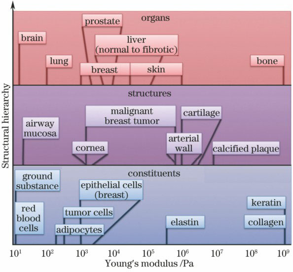
图 2. 一种典型的激光散斑生物组织黏弹性测量系统示意图
Fig. 2. System diagram of a typical viscoelastic measurement of biological tissues based on laser speckle

图 3. 根据散斑衬比变化测杨氏模量。(a)测量原理;(b)速度计算方法;(c)有硬块仿体的杨氏模量[38]
Fig. 3. Young's modulus measurement based on speckle contrast difference. (a) Measurement principle; (b) velocity calculation method; (c) Young's modulus of phantom with an inclusion

图 4. (a)人的血液样本凝固过程(第0,6,10,12 min)的散斑光强自相关曲线;(b)人的血液样本凝固过程中散斑自相关时间常数和黏弹性模量的变化
Fig. 4. (a) Speckle intensity autocorrelation curves measured during coagulation (at 0, 6, 10, 12 min) of a human blood sample; (b) changes of speckle autocorrelation time constant and viscoelasticity modulus during coagulation process of a human blood sample


陈肖, 陆锦玲, 李鹏程. 生物组织黏弹性激光散斑检测方法研究进展[J]. 中国激光, 2018, 45(2): 0207005. Chen Xiao, Lu Jinling, Li Pengcheng. Viscoelasticity Measurement of Biological Tissues Using Laser Speckle Techniques: a Review[J]. Chinese Journal of Lasers, 2018, 45(2): 0207005.


 PDF全文
PDF全文

