激光与光电子学进展, 2018, 55 (12): 120901, 网络出版: 2019-08-01
定量干涉显微流式细胞仪的研究与设计  下载: 1139次
Research and Design of Quantitative Interferometric Microscopic Cytometer
图 & 表
图 1. (a)~(d)视场扫描时不同位置上的血红细胞;(e)~(h)对应(a)~(d)中白色虚线框中的亚视场干涉图
Fig. 1. (a)-(d) Red blood cells at different positions during field of view scanning; (e)-(h) interferograms in sub-field of view corresponding to those in dashed boxes of (a)-(d)
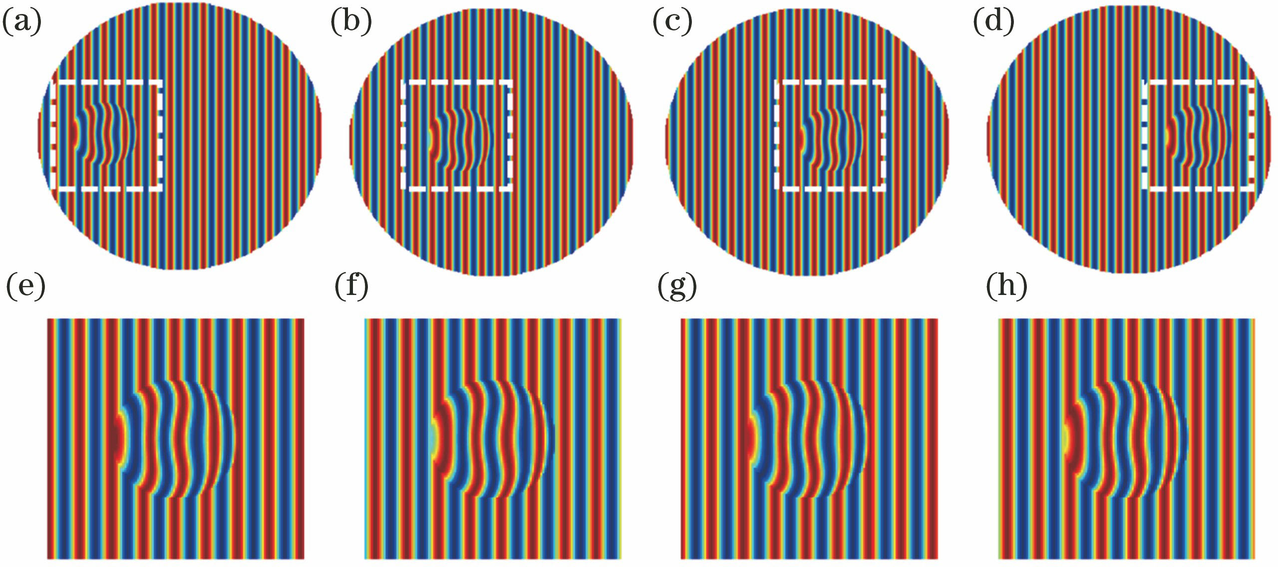
图 2. 血红细胞模型定量相位分布。(a)根据(2)式得到的血红细胞模型定量相位分布;(b)传统的基于快速傅里叶变换相位恢复算法得到的血红细胞模型定量相位分布;(c)传统的基于希尔伯特变换相位恢复算法得到的血红细胞模型定量相位分布;(d)根据扩展主程序分析相位恢复算法得到的血红细胞模型定量相位分布;(e)含有噪声的干涉图;(f)噪声情况下,根据扩展主程序分析相位恢复算法得到的血红细胞模型定量相位分布
Fig. 2. Quantitative phase distributions of red blood cell model. (a) Quantitative phase distribution of red blood cell model obtained according to equation (2); (b) quantitative phase distribution recovered by traditional phase retrieval algorithm based on fast Fourier transform; (c) quantitative phase distribution recovered by traditional phase retrieval algorithm based on Hilbert transform; (d) quantitative phase distribution recovered by expanded principle component analysis phase retrieval algorith

图 3. 基于机械扫描的定量干涉显微流式细胞仪
Fig. 3. Scheme of quantitative interferometric microscopic cytometer based on mechanic field of view scanning

图 4. (a)基于扩展主程序分析相位恢复算法的定量干涉显微流式细胞仪采集的干涉图;(b)相位恢复后的血红细胞定量相位分布
Fig. 4. (a) Interferograms captured by quantitative interferometric microscopic cytometer with phase retrieval algorithm based on expanded principle component analysis; (b) quantitative phase distribution of red blood cell after phase retrieval

图 5. (a)(b)视场扫描时在不同位置的血红细胞;(c)(d)对应(a)(b)中白色虚线的亚视场干涉图;(e)基于正则化光学流场相位恢复算法得到的被测样品相位;(f)噪声情况下,基于正则化光学流场相位恢复算法得到的血红细胞模型定量相位分布
Fig. 5. (a)(b) Red blood cells at different positions during field of view scanning; (c)(d) interferograms in sub-field of view corresponding to those in dashed boxes of (a) and (b); (e) phase of measured sample recovered by phase retrieval algorithm based on regularized optical flowing; (f) quantitative phase distribution of red blood cell model recovered by phase retrieval algorithm based on regularized optical flowing with noise

图 6. (a)基于正则化光学流场相位恢复算法的定量干涉显微流式细胞仪的操作步骤;(b)同一批次血红细胞的微分干涉相衬显微结果;(c)~(e)基于正则化光学流场相位恢复算法计算得到的被测样品的定量相位分布
Fig. 6. (a) Processing steps of quantitative interferometric microscopic cytometer with phase retrieval algorithm based on regularized optical flowing; (b) the same batch of red blood cells at differential interferometric contrast microscopy; (c)-(e) quantitative phase distributions of the measured sample recovered by phase retrieval algorithm based on regularized optical flowing

图 7. 重力驱动定量干涉显微流式细胞仪示意图
Fig. 7. Schematic of gravity-driven quantitative interferometric microscopic cytometer

图 8. 重力驱动的定量干涉显微流式细胞仪实现细胞大通量高速检测的流程
Fig. 8. Procedure of high-throughput and high-speed cell detection with gravity-driven quantitative interferometric microscopic cytometer

图 9. (a)微分干涉相衬显微镜下的同批次血红细胞样品;(b)~(f)重力驱动的定量干涉显微流式细胞仪得到的不同血红细胞定量相位分布
Fig. 9. (a) Same batch of red blood cell samples at differential interferometric contrast microscopy; (b)-(f) quantitative phase distributions of different red blood cells obtained by gravity-driven quantitative interferometric microscopic cytometer


表 1不同定量干涉显微流式细胞仪的对比
Table1. Comparison among different quantitative interferometric microscopic cytometers
|
闫克丁, 薛亮, 黄华川, 王绶玙. 定量干涉显微流式细胞仪的研究与设计[J]. 激光与光电子学进展, 2018, 55(12): 120901. Keding Yan, Liang Xue, Huachuan Huang, Shouyu Wang. Research and Design of Quantitative Interferometric Microscopic Cytometer[J]. Laser & Optoelectronics Progress, 2018, 55(12): 120901.


 PDF全文
PDF全文

