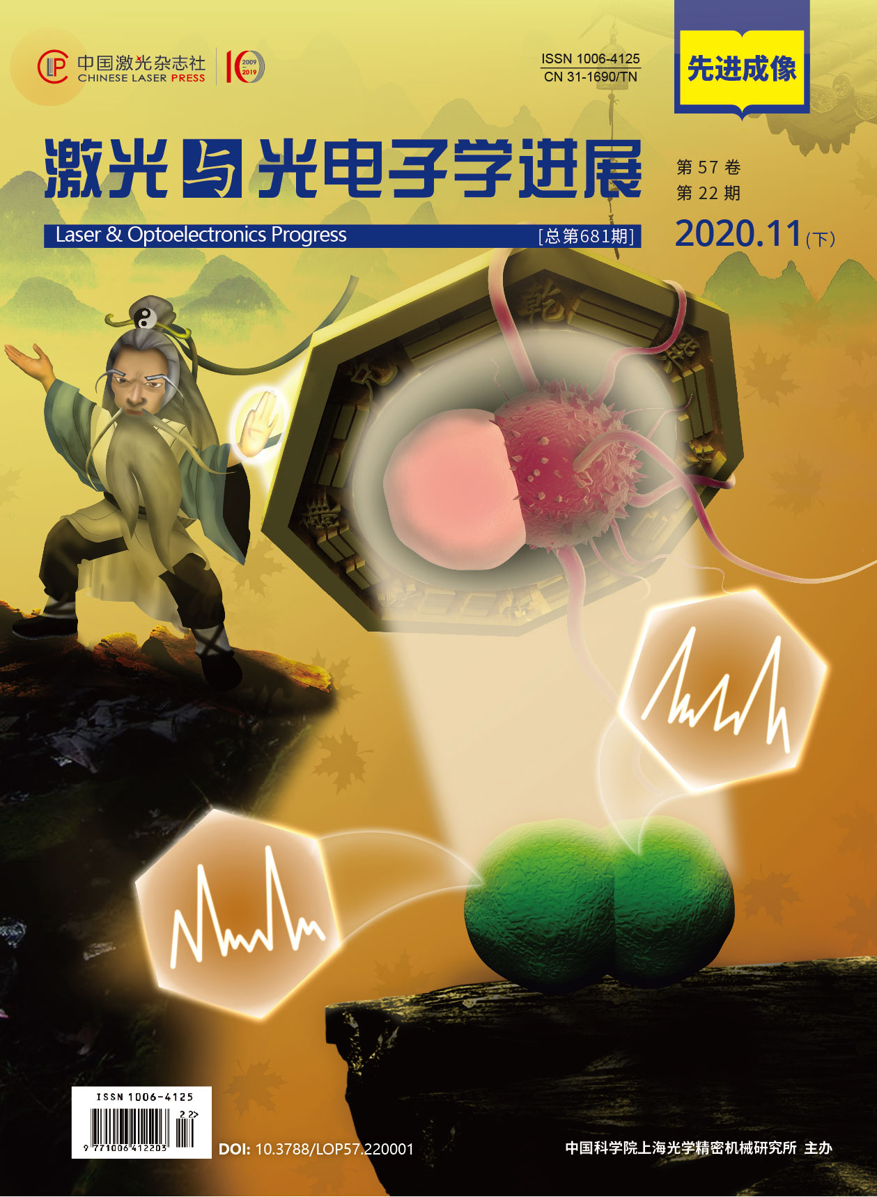
基于改进ResNeXt的乳腺癌组织病理学图像分类  下载: 1049次
牛学猛, 吕晓琪, 谷宇, 张宝华, 张明, 任国印, 李菁. 基于改进ResNeXt的乳腺癌组织病理学图像分类[J]. 激光与光电子学进展, 2020, 57(22): 221021.
Xuemeng Niu, Xiaoqi Lü, Yu Gu, Baohua Zhang, Ming Zhang, Guoyin Ren, Jing Li. Breast Cancer Histopathological Image Classification Based on Improved ResNeXt[J]. Laser & Optoelectronics Progress, 2020, 57(22): 221021.
[2] Spanhol FA, Oliveira LS, PetitjeanC, et al. Breast cancer histopathological image classification using convolutional neural networks[C]//2016 International Joint Conference on Neural Networks (IJCNN), July 24-29, 2016, Vancouver, BC, Canada. New York: IEEE, 2016: 2560- 2567.
[4] GolatkarA, AnandD, SethiA. Classification of breast cancer histology using deep learning[EB/OL]. [2020-03-23].https://arxiv.org/abs/1802. 08080.
[5] RakhlinA, ShvetsA, IglovikovV, et al. Deep convolutional neural networks for breast cancer histology image analysis[M] //Rakhlin A, Shvets A, Iglovikov V, et al. Image Analysis and Recognition. ICIAR 2018. Lecture Notes in Computer Science. Cham: Springer, 2018, 10882: 737- 744.
[6] KonéI, BoulmaneL. Hierarchical ResNeXt models for breast cancer histology image classification[M] //Campilho A, Karray F, ter Haar Romeny B. et al. Image Analysis and Recognition. ICIAR 2018. Lecture Notes in Computer Science. Cham: Springer, 2018, 10882: 796- 803.
[7] NazeriK, AminpourA, EbrahimiM. Two-stage convolutional neural network for breast cancer histology image classification[EB/OL]. [2020-03-21].https://arxiv.org/abs/1803. 04054.
[8] Wang Z H, You K Y, Xu J J, et al. Consensus design for continuous-time multi-agent systems with communication delay[J]. Journal of Systems Science and Complexity, 2014, 27(4): 701-711.
[10] Gu Y, Lu X Q, Yang L D, et al. Automatic lung nodule detection using a 3D deep convolutional neural network combined with a multi-scale prediction strategy in chest CTs[J]. Computers in Biology and Medicine, 2018, 103: 220-231.
[11] 孟婷, 刘宇航, 张凯昱. 一种基于增强卷积神经网络的病理图像诊断算法[J]. 激光与光电子学进展, 2019, 56(8): 081001.
[12] 李素梅, 雷国庆, 范如. 基于双通道卷积神经网络的深度图超分辨研究[J]. 光学学报, 2018, 38(10): 1010002.
[13] Xie SN, GirshickR, DollárP, et al. Aggregated residual transformations for deep neural networks[C]//2017 IEEE Conference on Computer Vision and Pattern Recognition (CVPR), July 21-26, 2017, Honolulu, HI, USA. New York: IEEE, 2017: 5987- 5995.
[14] Chen YP, Fan HQ, XuB, et al. Drop an octave: reducing spatial redundancy in convolutional neural networks with octave convolution[C]//2019 IEEE/CVF International Conference on Computer Vision (ICCV), October 27-November 2, 2019, Seoul, Korea (South). New York: IEEE, 2019: 3434- 3443.
[15] SinghP, Verma VK, RaiP, et al. HetConv: heterogeneous kernel-based convolutions for deep CNNs[C]//2019 IEEE/CVF Conference on Computer Vision and Pattern Recognition (CVPR), June 15-20, 2019, Long Beach, CA, USA. New York: IEEE, 2019: 4830- 4839.
[16] IoffeS, SzegedyC. Batch normalization: accelerating deep network training by reducing internal covariate shift[EB/OL]. [2020-03-23].https://arxiv.org/abs/1502. 03167.
[18] He KM, Zhang XY, Ren SQ, et al. Deep residual learning for image recognition[C]//2016 IEEE Conference on Computer Vision and Pattern Recognition (CVPR), June 27-30, 2016, Las Vegas, NV, USA. New York: IEEE, 2016: 770- 778.
[19] LinM, ChenQ, Yan SC, et al. Network in network[EB/OL]. [2020-03-25].https://arxiv.org/abs/1312. 4400.
[20] Srivastava N. Improving neural networks with dropout[J]. University of Toronto, 2013, 53(9): 1689-1699.
[23] Aresta G, Araújo T, Kwok S, et al. BACH: grand challenge on breast cancer histology images[J]. Medical Image Analysis, 2019, 56: 122-139.
[24] Gu Y, Lu X Q, Zhang B H, et al. Automatic lung nodule detection using multi-scale dot nodule-enhancement filter and weighted support vector machines in chest computed tomography[J]. PLOS One, 2019, 14(1): e0210551.
[25] 郭琳琳, 李岳楠. 基于专家乘积系统的组织病理图像分类算法[J]. 激光与光电子学进展, 2018, 55(2): 021008.
[26] Khan S, Islam N, Jan Z, et al. A novel deep learning based framework for the detection and classification of breast cancer using transfer learning[J]. Pattern Recognition Letters, 2019, 125: 1-6.
[27] 谷宇, 吕晓琪, 吴凉, 等. 基于NSCT和CLAHE的乳腺钼靶X线图像微钙化点增强方法[J]. 光学技术, 2018, 44(1): 6-12.
[28] GuptaV, BhavsarA. Breast cancer histopathological image classification: is magnification important?[C]//2017 IEEE Conference on Computer Vision and Pattern Recognition Workshops (CVPRW), July 21-26, 2017, Honolulu, HI, USA. New York: IEEE, 2017: 769- 776.
[29] Bardou D, Zhang K, Ahmad S M. Classification of breast cancer based on histology images using convolutional neural networks[J]. IEEE Access, 2018, 6: 24680-24693.
[30] 谷宇, 吕晓琪, 赵瑛, 等. 基于PSO-SVM的乳腺肿瘤辅助诊断研究[J]. 计算机仿真, 2015, 32(5): 344-349.
Gu Y, Lü X Q, Zhao Y, et al. Research on computer-aided diagnosis of breast tumors based on PSO-SVM[J]. Computer Simulation, 2015, 32(5): 344-349.
[31] 何雪英, 韩忠义, 魏本征. 基于深度学习的乳腺癌病理图像自动分类[J]. 计算机工程与应用, 2018, 54(12): 121-125.
He X Y, Han Z Y, Wei B Z. Breast cancer histopathological image auto-classification using deep learning[J]. Computer Engineering and Applications, 2018, 54(12): 121-125.
牛学猛, 吕晓琪, 谷宇, 张宝华, 张明, 任国印, 李菁. 基于改进ResNeXt的乳腺癌组织病理学图像分类[J]. 激光与光电子学进展, 2020, 57(22): 221021. Xuemeng Niu, Xiaoqi Lü, Yu Gu, Baohua Zhang, Ming Zhang, Guoyin Ren, Jing Li. Breast Cancer Histopathological Image Classification Based on Improved ResNeXt[J]. Laser & Optoelectronics Progress, 2020, 57(22): 221021.


