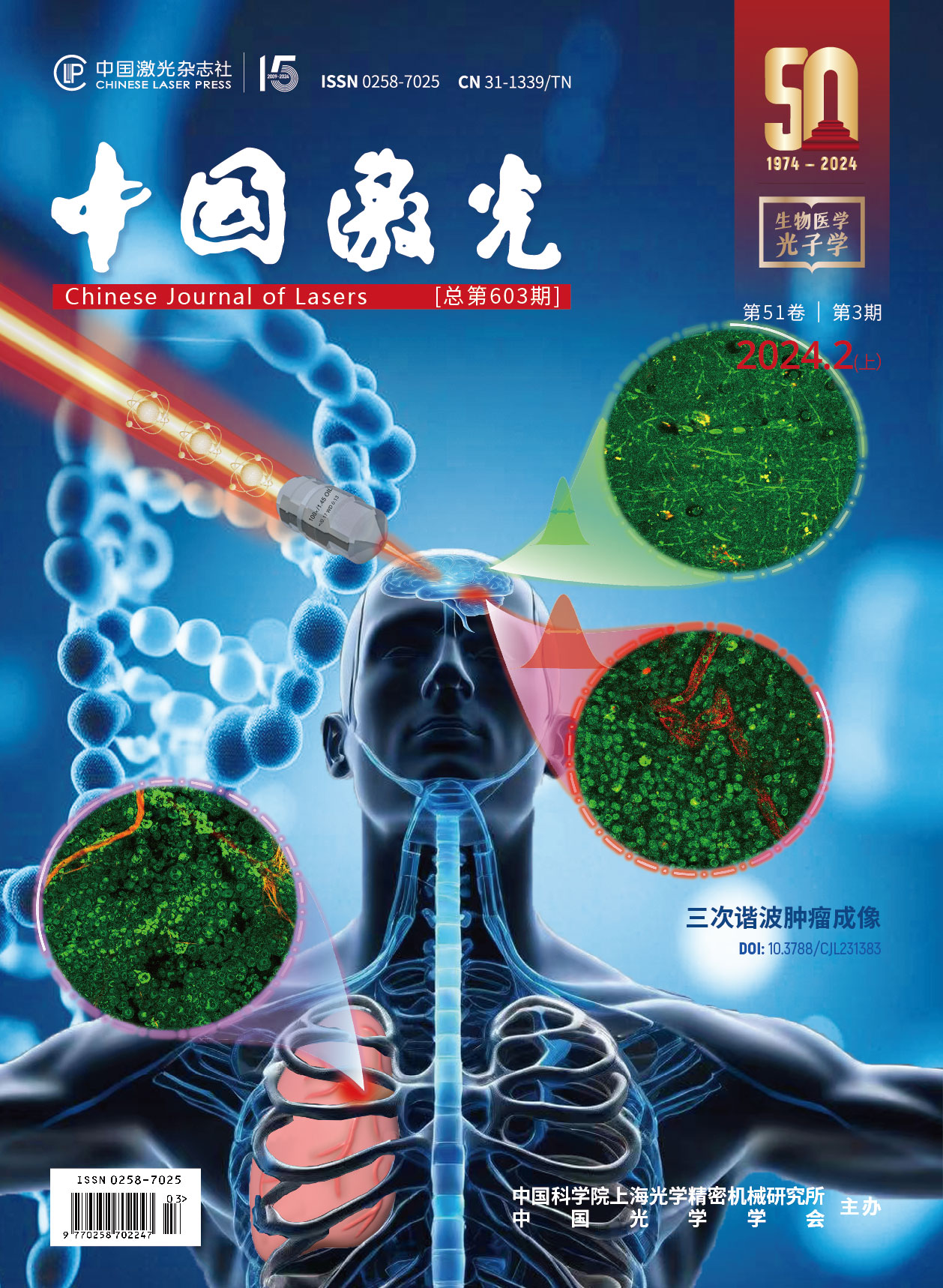
基于MC模型和Nelder‑Mead单纯形算法的时域组织光谱学
Changes in optical parameters can reflect the physiological status of biological tissue and constitute a fundamental and important topic in the field of near-infrared spectroscopy. Compared with the continuous-wave and frequency-domain measurement methods, the time-domain measurement method has the best performance in distinguishing and separating absorption and scattering coefficients, particularly in single-point measurement scenarios. Consequently, the time-domain measurement method is more commonly used to measure changes in optical parameters, also known as time-resolved spectroscopy. Currently, the biological tissue model used for time-resolved spectroscopy inversion schemes often assumes that the biological tissue is a single-layer biological tissue model, which hypothesizes that the optical properties are identical throughout the tissue. Although the single-layer biological tissue model simplifies the complexity of light propagation models and inversion algorithms, it is not very suitable for representing the structure of most human biological tissues; biological tissues at different depths exhibit a layered structure owing to variations in structure and function. Considering the high computational complexity and marginal improvement in accuracy associated with multilayer models, recent research has increasingly focused on the double-layer biological tissue model.
Currently, the commonly used double-layer biological tissue model parameter inversion methods face several challenges. First, they require a substantial amount of experimental data from multiple sources and detector separation, resulting in extended overall measurement times. Second, considerable time and effort are required to construct precise databases. Third, the need for iterative differentiation leads to prolonged computation time and lower accuracy. Finally, these methods struggle to handle complex scenarios, such as those in which both layers of tissue parameters are entirely unknown. To address these issues, this study introduces the Nelder-Mead simplex algorithm for the first time into the framework of Monte Carlo model-based tissue optical parameter inversion, developing a time-domain Monte Carlo supported Nelder–Mead simplex (MC-NMS) inversion algorithm.
The Monte Carlo model can use simulations to customize the optical parameters based on the structural characteristics of layered tissues, which are often employed as transport models for double-layer biological tissue model parameter inversion. This study introduces the Nelder–Mead simplex algorithm into Monte Carlo-based tissue optical parameter inversion for the first time. By utilizing only two source and detector separation time-domain diffuse reflectance data, the need to construct extensive databases in advance is eliminated. Initially, we empirically set the parameter values to find a feasible solution within the feasible region. The variables were incrementally adjusted through a cyclic iterative approach involving the establishment of different base vectors. Through matrix linear transformations, the optimal value of the objective function was determined using a heuristic search method that obviated the need for differentiation. Ultimately, this approach achieves a high-fidelity inversion of the optical parameters in layered tissues under complex conditions.
The numerical simulation experiments demonstrate that in the single-layer tissue optical parameter inversion application scenario, the proposed MC-NMS method yields significantly superior results compared to the TDIA and SDIA methods (Fig.3). Additionally, when the source-detector separation is set to 3 mm, the MC-NMS method yields the best results. (Fig.4). For double-layer biological tissue model optical parameter inversion, the results reveal that changing the upper-layer tissue thickness also requires different optimal source–detector separations (Fig.5, Fig.6). Moreover, the inversion errors obtained using the MC-NMS method are lower than those obtained using the TDIA and SDIA methods. Experimental validation was conducted using a multi-wavelength and multi-source-detector separation time-resolved measurement system based on time-correlated single-photon counting technology developed by our research group. Data were computed using the 25% rising edge to 20% falling edge time channels of the measured time-point spread function curves. The results indicate that the MC-NMS method achieves inversion errors of 11.64% and 0.89% in the single-layer biological tissue model for
The results of the liquid phantom experiments for the single-layer biological tissue and double-layer biological tissue models consistently demonstrate that the MC-NMS method outperforms other approaches. Notably, when the
张童, 刘东远, 高峰. 基于MC模型和Nelder‑Mead单纯形算法的时域组织光谱学[J]. 中国激光, 2024, 51(3): 0307203. Tong Zhang, Dongyuan Liu, Feng Gao. Time‑Resolved Spectroscopy Based on Monte Carlo Model and Nelder‑Mead Simplex Algorithm[J]. Chinese Journal of Lasers, 2024, 51(3): 0307203.


