中国激光, 2017, 44 (1): 0102005, 网络出版: 2017-01-10
激光微加工技术制备生物医用器械的现状与进展  下载: 2434次
Laser Microfabrication of Biomedical Devices
图 & 表
图 1. (a) 利用激光技术在单晶Si、PDMS、PLGA和ORMOCER表面制备的微结构;(b) NIH/3T3活细胞(绿色)和死细胞(黄-红色)在PDMS和PLGA结构表面的荧光显微图;(c) PC12活细胞(绿色)和死细胞(黄-红色)在PDMS和PLGA结构表面的荧光显微图
Fig. 1. (a) Laser created microstructures on the surface of single crystal silicon, PDMS, PLGA and ORMOCER; (b) fluorescence microscopy images of live (green) and dead (yellow-red) NIH/3T3 cells cultured on PDMS and PLGA micro-replicas; (c) fluorescence microscopy images of live (green) and dead (yellow-red) PC12 cells cultured on PDMS and PLGA micro-replicas
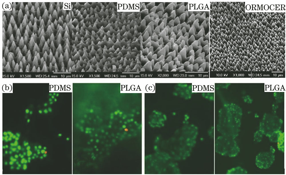
图 2. (a) 飞秒激光在Ti6Al4V合金表面加工的微纳结构;(b) 细胞在抛光表面及A、B、C 3种结构表面扩展面积随时间的变化;(c) 细胞在抛光表面及A、B、C 3种结构表面培养12天时脂肪形成表达基因的相对值
Fig. 2. (a) fs-laser created micro/nano structures on Ti6Al4V surface; (b) area of cell spreading on the polished surface and surfaces with textures A, B or C versus time; (c) relative value of MSCs adipogenic genes expression on the polished surface and surfaces with textures A, B or C after 12 d

图 3. (a) 激光直写技术在Ti6Al4V表面制得的微纳结构;(b) hMSCs在A、B、C 3种微纳结构表面的形状
Fig. 3. (a) Micro/nano structures on Ti6Al4V surface fabricated via laser direct writing method; (b) hMSCs cell shape on the surface with textures A, B, C

图 4. (a) 经激光加工和酸洗在TC4表面产生的微纳结构;(b) 成骨细胞在TC4抛光表面(1)、微结构表面(2)、微纳结构表面(3)培养4 h后的黏附率
Fig. 4. (a) Micro/nano structures on TC4 surface created via laser and pickling process; (b) adhesion rates of osteoblasts after culturing for 4 h on TC4 samples with polished surface, microstructure surface and micro-nanostructure surface

图 5. (a) 激光在PDMS和铂箔材料上加工的可移植高密度微电极阵列;(b) L929细胞在PDMS、铂箔和激光制备的微电极阵列结构上的相对增殖率(计算范围为细胞培养时间t=4 h至t=48 h)
Fig. 5. (a) Implantable high-density microelectrode array fabricated by laser-structuring of Pt foil and PDMS; (b) proliferation of L929 cells in direct contact with PDMS, Pt foil and the laser fabricated microelectrode array (The values are the rate of increase in cell number between t=4 h and t=48 h)

图 6. 激光在PDMS和Pt材料上加工的(a)多层微电极阵列及(b)其表面显微图
Fig. 6. (a) Multi-layer micro-electrode array fabricated by laser-structuring of Pt foil and PDMS and (b) its surface micrograph

图 7. 应用激光微加工技术分别在PDMS和PMMA上加工的微流道结构。(a) PDMS; (b) PMMA
Fig. 7. Microchannels fabricated on PDMS and PMMA via laser micromaching, respectively. (a) PDMS; (b) PMMA

图 8. 激光微加工制备的PPy基主动导管。(a) 四电极导管设计结构;(b) 激光微加工技术制备的四电极导管扫描电镜图;(c) 导管一端的PPy弯曲运动
Fig. 8. Laser fabricated PPy-based active catheter. (a) Designed model of four-electrode PPy-based active catheter; (b) scanning electron microscope photograph of four-electrode PPy-based active catheter fabricated via laser micromachining; (c) bend motion of PPy actuators strips on one end of the catheter

图 9. 在3D微纳纤维结构上制得的(a)孔隙阵列结构与(b)光学显微结构图;(c)激光微加工孔隙阵列结构表面的细胞分布共聚焦显微图像与(d)快速成型支架结构表面细胞分布SEM图,其中图(c)中蓝色代表细胞核,红色代表F-肌动蛋白
Fig. 9. (a) Porous arrays and (b) its optical microstructures on 3D micro/nanofibrous structure; (c) confocal microscope image of cell distributions on porous structures fabricated via laser micromachining (the blue and red colours indicate nuclei and F-actin, respectively ) and (d) SEM image of cell distributions on scaffold fabricated via laser rapid prototyping

图 10. (a)激光切割316L不锈钢血管支架与(b)局部结构的SEM图
Fig. 10. SEM photographs of (a) 316L stainless steel stent and (b) its local stent structure

图 11. 飞秒激光切割三角切口结构的(a)PLLA薄片和(b)局部结构
Fig. 11. (a) PLLA sheet with triangular cut-outs fabricated via fs-laser and (b) its zoomed local structure


图 14. (a) 利用SLS技术制备的PVDF支架结构;(b) PVDF支架在模拟体液中浸泡后的余重百分比随浸泡时间的变化曲线;(c) MG63在PVDF支架上培养1、3、5天后(从左至右)的生物活性
Fig. 14. (a) PVDF scaffold fabricated by SLS; (b) remaining weight percentage of PVDF scaffolds after immersed in simulated body fluid for different days; (c) bioactivity of MG63 cells after 1, 3, 5 d cultured on PVDF scaffolds (from left to right)


图 16. MG63和hBMSCs在不同晶粒度支架上的SEM形貌图。MG63:(a) 1.32 μm;(b) 0.71 μm;(c) 0.21 μm;hBMSCs:(d) 1.32 μm;(e) 0.71 μm;(f) 0.21 μm
Fig. 16. SEM morphology of MG63 and hBMSCs cultured onto scaffolds with different grain sizes. MG63 cells:(a) 1.32 μm; (b) 0.71 μm; (c) 0.21 μm; hBMSCs cells: (d) 1.32 μm; (e)0.71 μm; (f) 0.21 μm

图 17. 利用SLM技术制备的不同尺寸与结构的骨支架结构
Fig. 17. SLM fabricated bone scaffold with different sizes and unit structures


表 1支架的溶血实验结果[56]
Table1. Hemolysis experimental results of stent[56]
|
卢立斌, 王海鹏, 管迎春, 周伟. 激光微加工技术制备生物医用器械的现状与进展[J]. 中国激光, 2017, 44(1): 0102005. Lu Libin, Wang Haipeng, Guan Yingchun, Zhou Wei. Laser Microfabrication of Biomedical Devices[J]. Chinese Journal of Lasers, 2017, 44(1): 0102005.


 PDF全文
PDF全文

