中国激光, 2018, 45 (3): 0307008, 网络出版: 2018-03-06
光声显微成像技术研究进展及其应用  下载: 2759次特邀综述
Progress and Application of Photoacoustic Microscopy Technique
图 & 表
图 1. (a)声学分辨率光声显微镜[1];(b)光学分辨率显微镜[7];(c)超分辨率光声显微镜[8];(d)基于中空超声换能器的光声显微镜[11]
Fig. 1. (a) Acoustic-resolutionphotoacoustic microscopy[1]; (b) optical-resolution photoacoustic microscopy[7]; (c) super-resolution photoacoustic microscopy[8]; (d) hollow-focused ultrasonic transducer based photoacoustic microscopy[11]
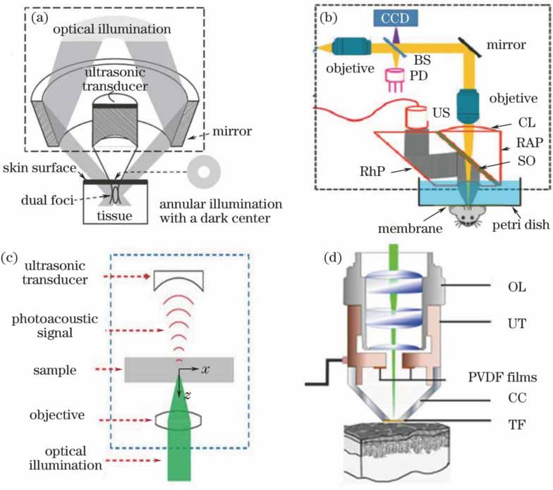
图 2. (a)血管网络光声图像;(b)血氧饱和度图像;(c)(d)血流速度成像[31]
Fig. 2. (a) Photoacoustic image of blood vessel; (b) image of oxygen saturation; (c)(d) images of blood velocity[31]

图 3. (a)不同浓度琼脂样品与(b)斑块样品[35]的光声粘弹图像
Fig. 3. Photoacoustic viscoelasticity images of (a) phantoms containing various concentrations of lipid and (b) atherosclerotic plaque sample[35]

图 4. (a)细胞核、(b)红细胞、(c)视网膜血管网络及色素层、(d)伊文思蓝染料增强后的血管网络、(e)金纳米粒子靶向B16黑色素瘤、(f) iRFP荧光蛋白过表达肿瘤[37]的光声图像
Fig. 4. Photoacoustic images of (a) cell nuclei, (b) red blood cells, (c) retinal vessels and retinal pigment epithelium layer, (d) microvasculature enhanced by Evans blue dye, (e) B16 melanoma labeled with targeted gold nanocages, (f) tumor expressed by near-infrared fluorescent protein iRFP[37]

图 5. (a)基于法布里珀罗标准具的光声成像系统[45];(b)基于聚合物微环谐振器的光声成像系统[58];(c)基于低相干迈克耳孙干涉仪的光声成像系统[63]
Fig. 5. (a) Fabry-Perot polymer film etalon ultrasound sensor based photoacoustic imaging system[45]; (b) polymer microring resonators based photoacoustic imaging system[58]; (c) low coherence Michelson interferometer based photoacoustic imaging system[63]

图 6. 多模态系统成像活体老鼠耳朵[70]。(a)光声最大值投影;(b) OCT最大值投影;(c)荧光最大值投影, 插图为体视显微镜下获取的小鼠耳朵的荧光图像;(d)光声/OCT/荧光融合图像;(e)对应(a)中1处的光声/OCT断层图像;(f)对应(a)中2处的光声/OCT断层图像
Fig. 6. In vivo imaging of mouse ear using multi-modal imaging system[70]. Images of (a) AOPAM maximum amplitude projection, (b) OCT maximum amplitude projection, and (c) FLM maximum amplitude projection; (d) image fused AOPAM, OCT and FLM; (e) AOPAM, OCT and fused AOPAM-OCT B-scan images of white dotted line 1 in (a); (f) AOPAM, OCT and fused AOPAM-OCT B-scan images of white dotted line 2 in (a); inset in (c) is fluorescence image of mouse ears obtained under a stereomicroscope

图 7. (a)合成孔径聚焦算法原理;(b)相干因子加权说明[75]
Fig. 7. (a) Schematic of virtual-detector concept; (b) illustration of coherent factor weighting[75]

图 8. (a)小鼠耳朵血管网络[7]与(b)脑皮层血管网络、血氧饱和度[84]的光声图像
Fig. 8. Photoacoustic images of (a) mouse ear vasculature[7] and (b) mouse brain vasculature and oxygen saturation[84]

图 9. 全光光声(AOPA)和OCT系统的活体成像结果。(a)正常、(b)黑色素瘤、(c)基底细胞癌小鼠耳朵的光声显微图像;(d)~(f)白色点线对应光声OCT的B扫光声图像;(g)~(i)对应的H&E染色组织切片[71]
Fig. 9. In vivo imaging results by all-optical photoacoustic (AOPA) and OCT system. AOPA maximum amplitude projection images of (a) normal, (b) BCC, and (c) MM mouse ear; (d)-(f) OCT B-scan images for white dotted line in (a)-(c); (g)-(i) imaging with H&E staining histopathology[71]


陈重江, 杨思华, 邢达. 光声显微成像技术研究进展及其应用[J]. 中国激光, 2018, 45(3): 0307008. Chen Zhongjiang, Yang Sihua, Xing Da. Progress and Application of Photoacoustic Microscopy Technique[J]. Chinese Journal of Lasers, 2018, 45(3): 0307008.


 PDF全文
PDF全文

