激光与光电子学进展, 2019, 56 (7): 070005, 网络出版: 2019-07-30
无标记显微成像技术的研究进展  下载: 2380次
Research Progress on Label-Free Microscopic Imaging Technology
图 & 表
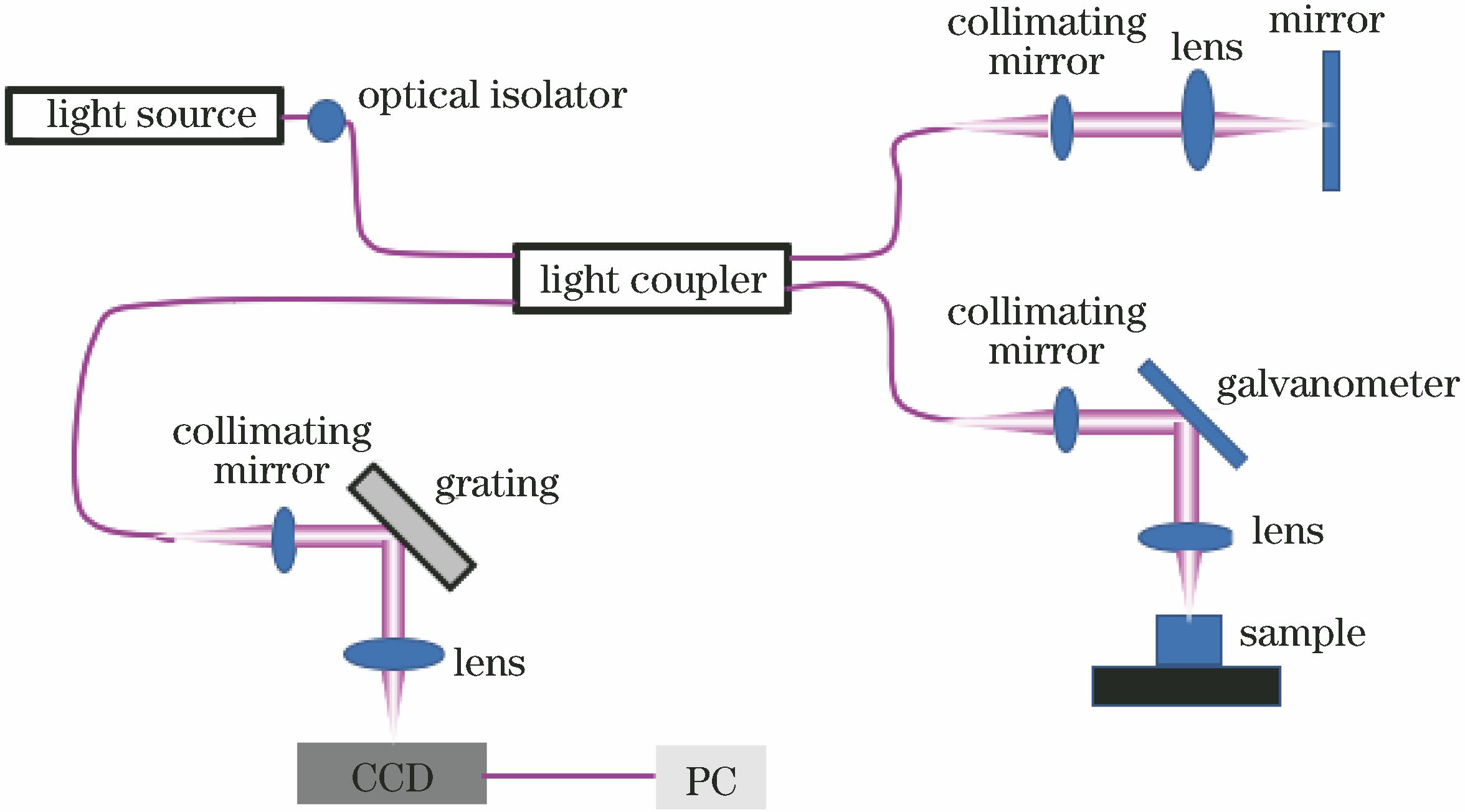
图 2. 人眼视网膜Angio-OCT图像。(a)超大视场(12 mm×16 mm)人眼视网膜Angio-OCT图像;(b)视神经乳头图像;(c)中心凹图像;(d)颞区图像(视场为2.0 mm×2.4 mm)[9]
Fig. 2. Angio-OCT images of human retina. (a) Large field of view (12 mm×16 mm) for Angio-OCT image of human retina; (b) optic nerve papilla image; (c) fovea image; (d) temporal region image (field of view is 2.0 mm×2.4 mm)[9]

图 3. 手掌表皮MIP图。(a)表皮-真皮交界处MIP;(b)表皮-真皮交界处的小血管和毛细血管袢MIP;(c)水平神经丛下方的大血管MIP[21]
Fig. 3. MIP of palm skin.(a)MIP of the epidermal-dermal junction; (b) MIP of the smaller vessels and captllary loops at the epidermal-dermal junction; (c) MIP of the bigger vesels under the horizontal plexus[21]

图 4. THG显微镜揭示的形态特征总结[29]
Fig. 4. Summary of morphological features revealed by THG microscopy[29]

图 5. 浸没式微球透镜的腺病毒簇成像结果。(a)腺病毒簇的高放大倍数SEM图像;(b)腺病毒簇的低放大倍率SMON图像(100 μm BaTiO3微球与503目标镜片偶联,NA=0.75);(c)腺病毒簇的更高放大倍数SEM图像;(d)腺病毒簇的较高放大倍数SMON图像(100 μm BaTiO3微球与803物镜耦合,NA=0.9)[37]
Fig. 5. Adenovirus clusters images of immersed microsphere lens. (a) High magnification SEM image of adenovirus clusters; (b) low magnification SMON image of adenovirus clusters (100 μm BaTiO3 microsphere coupled with a 503 objective lens, NA=0.75); (c) higher magnification SEM image of adenovirus clusters; (d) higher magnification SMON image of adenovirus clusters (100 μm BaTiO3 microsphere coupled with an 803 objective lens, NA=0.9)[37]

图 6. 活体小鼠胚胎的心脏血管内的血流动力学多普勒OCT成像
Fig. 6. Doppler OCT imaging of hemodynamics inside heart tube of a live mouse embryo

图 7. 离体人脑组织的THG / SHG内窥显微镜检查。(a)神经元胞体组织;(b)灰质神经毡组织;(c)低细胞性白质区域;(d)高级别胶质瘤[55]
Fig. 7. THG/SHG endomicroscopy results of ex-vivo human brain tissues. (a) Neuronal somata tissue; (b) gray matter neuropil tissue; (c) low-cellularity white matter area; (d) high-grade glioma[55]

图 8. CARS显微镜追踪脂滴形成实验。(a)间充质干细胞分化成脂肪的过程;(b)使用Imaris软件包(Bitplane)描绘脂滴的体积变化;(c1)主受体脂滴的表面增加与熔合脂滴提供的净表面之间的比较;(c2)主受体脂滴的体积增加与所有熔合脂滴的净体积之间的比较[63]
Fig. 8. CARS microscope tracking lipid droplet formation experiment. (a) Progress of differentiation of mesenchymal stem cells into adipose; (b) volume change of the lipid droplet depicted by the Imaris software (Bitplane); (c1) comparison between the surface increase of the main acceptor lipid droplet and the net surface provided by the fusing lipid droplet; (c2) comparison between the volume increase of the main acceptor lipid droplet and the net volume of all fusing lipid droplets[63]

图 9. 多模态成像活体老鼠耳朵。(a)光声成像图像;(b) OCT图像;(c) FLM的荧光最大值投影(MAP);(d) OCT/PAI/FLM融合图像;(e)对应图9(a)中1处的OCT/PAI断层图像;(f)对应图9(a)中2处的OCT/PAI断层图像
Fig. 9. Multimodal imaging of living mouse ears. (a) image of PAI; (b) image of OCT;(c) MAP image of FLM; (d) fused image of PAI, OCT, and FLM; (e) OCT/PAI tomography image corresponding to 1 in Fig. 9 (a); (f) OCT/PAI tomography image corresponding to 2 in Fig. 9(a)[66]

张佳, 洪亮, 任升, 周非凡, 胡睿, 屈军乐, 刘丽炜. 无标记显微成像技术的研究进展[J]. 激光与光电子学进展, 2019, 56(7): 070005. Jia Zhang, Liang Hong, Sheng Ren, Feifan Zhou, Rui Hu, Junle Qu, Liwei Liu. Research Progress on Label-Free Microscopic Imaging Technology[J]. Laser & Optoelectronics Progress, 2019, 56(7): 070005.


 PDF全文
PDF全文

