中国激光, 2020, 47 (2): 0207005, 网络出版: 2020-02-21
相干拉曼散射显微技术及其在生物医学领域的应用  下载: 3089次特邀综述
Coherent Raman Scattering Microscopy Technique and Its Biomedical Applications
图 & 表
图 1. CARS和SRS过程能级图。(a) CARS能级图;(b) SRS能级图
Fig. 1. Energy level diagrams of CARS and SRS processes. (a) CARS energy level diagram; (b) SRS energy level diagram
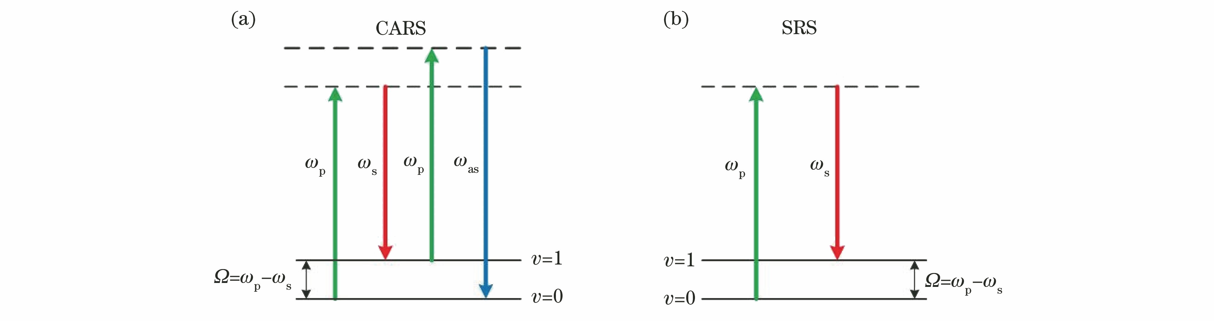

图 3. 用CARS显微镜检测到的富含脂质的前列腺循环肿瘤细胞(上排:前列腺循环肿瘤细胞表现出由细胞内脂质积累引起的强的CARS信号;下排:白细胞表现出主要来自细胞膜的弱的CARS信号)[36]
Fig. 3. Detection of lipid-rich prostate CTC with CARS microscopy. (Upper row: prostate CTC exhibits strong CARS signal arising from intracellular lipid accumulation; lower row: leukocytes exhibit weak CARS signal arising mainly from cellular membrane)[36]

图 4. 宽带CARS对小鼠胰管的三维组织成像[16]。(a)伪彩色图,蓝色为细胞核(785 cm-1),红色为胶原蛋白(855 cm-1),绿色为脂质和蛋白质的复合物(1665 cm-1),A为腺泡细胞,D为外分泌管,Ep为上皮细胞;(b)小鼠胰管的三维重建图
Fig. 4. Three-dimensional tissue imaging with wideband CARS for murine pancreatic ducts[16]. (a) Pseudo color chart, in which nuclei (785 cm-1), collagen (855 cm-1), and a composite of lipids and proteins (1665 cm-1) are highlighted in blue, red, and green, respectively. A represents acinar cell, D represents exocrine duct, and Ep represents epithelial cell; (b) three-dimensional reconstruction result of murine pancreatic

图 5. 利用标记和无标记成像方法对秀丽隐杆线虫体内脂肪成像的结果[48]。(a)采用苏丹黑、油红O和尼罗红对固定后的线虫进行染色的结果,以及分别采用尼罗红、BODIPY标记的脂肪酸喂养活线虫,并对脂肪进行标记成像的结果; (b)利用CARS和双光子激发荧光(TPEF)对中性脂质和自发荧光颗粒进行无标记成像的结果
Fig. 5. Labeled and label-free imaging for fat in C. elegans[48]. (a) Labeled imaging of fat using Sudan Black, Oil Red O, and Nile Red staining of fixed worms and Nile Red and BODIPY-labeled fatty acids fed to live worms; (b) label-free imaging results of neutral lipid species and autofluorescent gut granules using CARS and TPEF

图 6. 具有高浓度药物的小鼠BaF3细胞的高光谱SRS成像[32]。高浓度伊马替尼(a)和尼罗替尼(b)在小鼠BaF3细胞内的无标记可视化成像,这与仅使用二甲基亚砜处理的对照细胞(c)不同;选定位置的SRS光谱(d)与溶液中两种SRS光谱(e)相似,但与细胞质区域(f)不同
Fig. 6. Hyperspectral SRS imaging of BaF3 cells in mouce with high concentration drug. Unlabeled visualization imaging of high concentrations of imatinib (a) and nilotinib (b) in mouse BaF3 cells, as distinct from control cells treated with dimethyl sulfoxide alone (c); SRS spectrum (d) of the selected location shows similarity to two SRS spectra (e) in solution, but it is different from the cytoplasmic region (f)

图 7. 小鼠大脑肿瘤的SRS成像[53]。(a)在皮层表面下方的肿瘤中,在对皮层表面成像时,SRS(左)和明场像(右)没有明显异常;(b)去除一部分皮质后可发现肿瘤区域,SRS(左)和明场像(右),能够看到肿瘤与正常大脑之间的界面
Fig. 7. SRS imaging of brain tumors in mice[53]. (a) In tumors below the cortical surface, there were no significant abnormalities in the SRS (left) and brightfield images (right) when imaging the cortical surface; (b) after removing a part of the cortex, tumor regions can be found, and interface between the tumor and the normal brain can be seen in SRS (left) and brightfield images(right)

图 8. 秀丽隐杆线虫的CARS和SRS成像[64]。(a)共振背景下秀丽隐杆线虫肠细胞核的CARS图像;(b)秀丽隐杆线虫肠细胞中的SRS图像;相同位置的秀丽隐杆线虫中的CH2在2845 cm-1处的CARS(c)和SRS(d)放大图像
Fig. 8. CARS and SRS imaging of in C. elegans. (a) CARS images of C. elegans intestinal cells in resonance background; (b) SRS images of C. elegans intestinal cells; CARS (c) and SRS (d) enlarged images of C. elegans CH2 at 2845 cm-1 in the same location

李姿霖, 李少伟, 张思鹭, 沈炳林, 屈军乐, 刘丽炜. 相干拉曼散射显微技术及其在生物医学领域的应用[J]. 中国激光, 2020, 47(2): 0207005. Li Zilin, Li Shaowei, Zhang Silu, Shen Binglin, Qu Junle, Liu Liwei. Coherent Raman Scattering Microscopy Technique and Its Biomedical Applications[J]. Chinese Journal of Lasers, 2020, 47(2): 0207005.


 PDF全文
PDF全文

