激光与光电子学进展, 2019, 56 (7): 070001, 网络出版: 2019-07-30
光声成像技术在早期癌症检测治疗中的潜在应用  下载: 1885次
Potential Applications of Photoacoustic Imaging in Early Cancer Diagnosis and Treatment
图 & 表
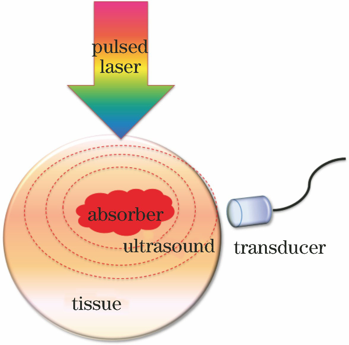
图 2. 光声成像系统。(a)透射式OR-PAM系统;(b)反射式OR-PAM系统;(c)暗场照明AR-PAM系统;(d)环形超声换能器阵列PACT系统;(e)线性超声换能器阵列PACT系统;(f)半球形超声换能器阵列PACT系统;(g)基于二维法布里-珀罗干涉仪声学传感器的PACT系统;(h)血管内侧视光声导管[46]
Fig. 2. Photoacoustic imaging systems. (a) Transmission-mode OR-PAM system; (b) reflection-mode OR-PAM system; (c) AR-PAM system with dark-field illumination; (d) PACT system with ring-shaped UTA; (e) PACT system with linear UTA; (f) PACT system with hemispherically shaped UTA; (g) PACT system based on 2D Fabry-Perot interferometer acoustic sensor; (h) side-viewing intravascular PA catheter[46]

图 3. 乳腺癌的光声成像[30]。(a)固定未处理的乳腺癌UV-PAM图;(b)经过切片和染色处理的乳腺组织与(a)中相同区域的H&E染色的组织学图像;(c)(d)对应(a)(b)中的红色虚线区域放大后的UV-PAM和H&E染色图像;(e)(f)对应(a)(b)中的黄色虚线区域放大后的UV-PAM和H&E图像;(g)放大(a)中橙色虚线区域的UV-PAM图像
Fig. 3. Photoacoustic imaging of breast tumor[30]. (a) UV-PAM image of fixed, unprocessed breast tumor; (b) H&E-stained histologic image of same area shown in Fig. (a) acquired after sectioning and staining breast tissue; (c)(d) zoomed UV-PAM and H&E-stained images of red dashed regions in Fig. (a) and (b), respectively; (e)(f) zoomed UV-PAM and H&E images of yellow dashed regions in Fig. (a) and (b), respectively; (g) zoomed UV-PAM image of orange dashed region in Fig. (a)


图 5. 光声成像测量得到的肿瘤内的血氧浓度。(a)两个单波长振幅PAR技术;(b)双波长振幅微分PAR成像技术;(c)两个单波长相位滤波微分PAR技术;(d)双波长相位滤波微分PAR技术[80]
Fig. 5. Oxygenation levels within tumor using photoacoustic imaging. (a) Two single-wavelength PAR amplitude technique; (b) two-wavelength differential PAR amplitude technique; (c) two single-wavelength phase-filtered differential PAR technique; (d) two-wavelength phase-filtered differential PAR technique[80]

图 6. 肿瘤的光声黏弹性成像。(a)肿瘤的光吸收图像;(b)肿瘤的黏弹性成像;(c)标记的肿瘤区域与正常组织的PA信号的平均幅度和相位延迟[91]
Fig. 6. Photoacoustic viscoelastic imaging of tumor. (a) Optical absorption image of tumor; (b) viscoelasticity image of tumor; (c) averaged amplitude and phase delay of PA signal from marked tumor region and normal tissue [91]

图 7. 前列腺肿瘤的光声功率谱成像[100]。(a)肿瘤和正常组织在感兴趣区域的超声成像;(b) PA图像与超声图像叠加后的中频带拟合结果;(c) PA图像与超声图像叠加后的斜率;(d) PA图像与超声图像叠加后的截距
Fig. 7. Photoacoustic power spectral imaging of prostate tumors[100]. (a) Ultrasonic imaging in region of interests for tumor and normal tissue; (b) mid-band fitting result after superposition of PA image and ultrasonic image; (c) slope after superposition of PA image and ultrasonic image; (d) intercept after superposition of PA image and ultrasonic image

吴华钦, 王昊宇, 谢文明, 李志芳, 吴淑莲, 李晖. 光声成像技术在早期癌症检测治疗中的潜在应用[J]. 激光与光电子学进展, 2019, 56(7): 070001. Huaqin Wu, Haoyu Wang, Wenming Xie, Zhifang Li, Shulian Wu, Hui Li. Potential Applications of Photoacoustic Imaging in Early Cancer Diagnosis and Treatment[J]. Laser & Optoelectronics Progress, 2019, 56(7): 070001.


 PDF全文
PDF全文

