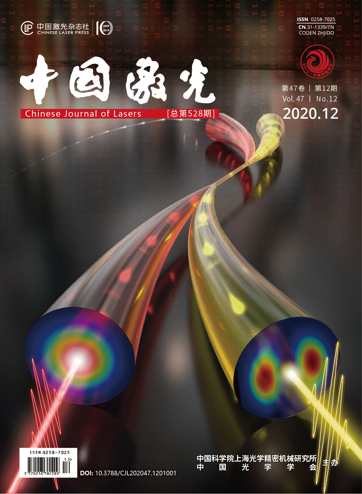
幼年到成年斑马鱼大脑的OCT活体三维可视化  下载: 1336次
毛广娟, 林燕萍, 陈婷如, 张艺晴, 邱婷, 蓝银涛, 向湘, 傅洪波, 张建. 幼年到成年斑马鱼大脑的OCT活体三维可视化[J]. 中国激光, 2020, 47(12): 1207002.
Mao Guangjuan, Lin Yanping, Chen Tingru, Zhang Yiqing, Qiu Ting, Lan Yintao, Xiang Xiang, Fu Hongbo, Zhang Jian. OCT in vivo Three-Dimensional Visualization of Zebrafish Brains from Juvenile to Adult[J]. Chinese Journal of Lasers, 2020, 47(12): 1207002.
[1] 李萍萍, 马涛, 张鑫, 等. 各国脑计划实施特点对我国脑科学创新的启示[J]. 同济大学学报(医学版), 2019, 40(4): 397-401.
Li P P, Ma T, Zhang X, et al. Enlightenment for China's brain science innovation from global brain projects[J]. Journal of Tongji University (Medical Science), 2019, 40(4): 397-401.
[5] 黄春念, 张晶晶. 模式动物斑马鱼在中枢神经系统疾病研究中的应用[J]. 中国实验动物学报, 2018, 26(3): 392-397.
Huang C N, Zhang J J. Research progress on the application of zebrafish in central nervous system diseases[J]. Acta Laboratorium Animalis Scientia Sinica, 2018, 26(3): 392-397.
[6] 蒋鹏翀, 魏巍, 胡芬, 等. 基于激光消融技术的斑马鱼条纹再生的研究[J]. 中国激光, 2018, 45(2): 0207024.
[7] 朱雨, 杨光, 李思黾, 等. 斑马鱼肌肉结构的定量偏振成像[J]. 光学学报, 2019, 39(8): 0811001.
[8] Randlett O, Wee C L, Naumann E A, et al. Whole-brain activity mapping onto a zebrafish brain atlas[J]. Nature Methods, 2015, 12(11): 1039-1046.
[9] 孙乐, 杜久林. 大脑神经联接图谱的研究进展[J]. 中国科学:生命科学, 2018, 48(3): 253-265.
Sun L, Du J L. Progress in brain neural connectomics[J]. Scientia Sinica (Vitae), 2018, 48(3): 253-265.
[12] Huang D, Swanson E A, Lin C P, et al. Optical coherence tomography[J]. Science, 1991, 254(5035): 1178-1181.
[13] Fercher A F, Drexler W, Hitzenberger C K, et al. Optical coherence tomography——principles and applications[J]. Reports on Progress in Physics, 2003, 66(2): 239-303.
[14] 韩涛, 邱建榕, 王迪, 等. 光学相干层析显微成像的技术与应用[J]. 中国激光, 2020, 47(2): 0207004.
[15] Yaqoob Z, Wu J G, Yang C. Spectral domain optical coherence tomography: a better OCT imaging strategy[J]. BioTechniques, 2005, 39(6S): S6-S13.
[16] 李培, 杨姗姗, 丁志华, 等. 傅里叶域光学相干层析成像技术的研究进展[J]. 中国激光, 2018, 45(2): 0207011.
[17] 魏波, 袁治灵, 唐志列. 基于光热光学相干层析技术的肿瘤组织三维成像[J]. 光学学报, 2020, 40(4): 0411002.
[18] 潘柳华, 张向阳, 李中梁, 等. 基于光声-光学相干层析成像的血流测量技术[J]. 中国激光, 2018, 45(6): 0607004.
[19] Jang I K, Bouma B E, Kang D H, et al. Visualization of coronary atherosclerotic plaques in patients using optical coherence tomography: comparison with intravascular ultrasound[J]. Journal of the American College of Cardiology, 2002, 39(4): 604-609.
[20] Nassif N, Cense B, Park B H, et al. In vivo high-resolution video-rate spectral-domain optical coherence tomography of the human retina and optic nerve[J]. Optics Express, 2004, 12(3): 367-376.
[21] Østby Y, Tamnes C K, Fjell A M, et al. Heterogeneity in subcortical brain development: a structural magnetic resonance imaging study of brain maturation from 8 to 30 years[J]. The Journal of Neuroscience, 2009, 29(38): 11772-11782.
[22] Divakar Rao K, Alex A, Verma Y, et al. Real-time in vivo imaging of adult zebrafish brain using optical coherence tomography[J]. Journal of Biophotonics, 2009, 2(5): 288-291.
[23] Zhang J, Ge W, Yuan Z. In vivo three-dimensional characterization of the adult zebrafish brain using a 1325nm spectral-domain optical coherence tomography system with the 27 frame/s video rate[J]. Biomedical Optics Express, 2015, 6(10): 3932-3940.
[25] Bai X, Qi Y M, Liang Y Z, et al. Photoacoustic computed tomography with lens-free focused fiber-laser ultrasound sensor[J]. Biomedical Optics Express, 2019, 10(5): 2504-2512.
[26] Seo E, Lim J, Seo S J, et al. Whole-body imaging of a hypercholesterolemic female zebrafish by using synchrotron X-ray micro-CT[J]. Zebrafish, 2015, 12(1): 11-20.
[27] Merrifield G D, Mullin J, Gallagher L, et al. Rapid and recoverable in vivo magnetic resonance imaging of the adult zebrafish at 7T[J]. Magnetic Resonance Imaging, 2017, 37: 9-15.
[28] Lin Y P, Chen T R, Mao G J, et al. Long-term and in vivo assessment of Aβ protein-induced brain atrophy in a zebrafish model by optical coherence tomography[J]. Journal of Biophotonics, 2020, 13(7): e202000067.
毛广娟, 林燕萍, 陈婷如, 张艺晴, 邱婷, 蓝银涛, 向湘, 傅洪波, 张建. 幼年到成年斑马鱼大脑的OCT活体三维可视化[J]. 中国激光, 2020, 47(12): 1207002. Mao Guangjuan, Lin Yanping, Chen Tingru, Zhang Yiqing, Qiu Ting, Lan Yintao, Xiang Xiang, Fu Hongbo, Zhang Jian. OCT in vivo Three-Dimensional Visualization of Zebrafish Brains from Juvenile to Adult[J]. Chinese Journal of Lasers, 2020, 47(12): 1207002.


