氧化石墨烯涂覆的空心微泡腔回音壁谐振模气体传感器【增强内容出版】
The trace gas direction holds practical significance in human health, industrial production safety, national defense, and other key fields. The optical fiber whisper gallery mode (WGM) sensors can achieve high sensitivity and resolution sensing measurement due to their strong light-matter interaction. However, the common silica material of WGM sensors is not sensitive to gases, which limits their applications in gas sensing. As a kind of two-dimensional material, graphene oxide (GO) not only has sound physical properties such as high mechanical strength and flexibility, but also features a significant surface volume ratio, efficient surface adsorption, low noise level, and stable chemical properties. Based on optical WGM excitation, the GO film is coated on a hollow microsphere cavity inwall to achieve gas sensing. The gas molecule adsorption on the GO will affect the effective refractive index of the overall microcavity structure and be reflected by the WGM shift. It is worth noting that the unique hollow structure of the microbubble is a natural fluid channel, which is very suitable for gas transportation. It is unnecessary to design a separate fluid channel or external gas chamber.
The investigation is based on the WGM sensor theory. The changed refractive index induced by gas molecular adsorption is analyzed. The sensors are fabricated by melt pressured rheological method and injection of GO dispersion. First, the performance of the GO-coated WGM gas sensor is investigated, and the changes in WGM resonance wavelength are observed by injecting gases with different concentrations into the sensor. Next, the gas sensing performance below 40×10-6 is elaborately investigated. The sensitivity and resolution of the sensor are obtained. Finally, the real-time response to 10×10-6-40×10-6 NH3 is demonstrated to show the sound recoverability, response, and recovery time.
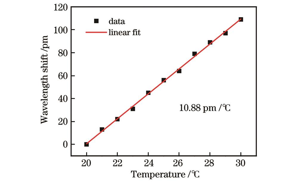
The designed GO-coated microbubble sensor exhibits deserved gas sensing performance. Fig. 4 shows the WGM spectrum of the structure with different gas concentrations. The resonance wavelength appears to be red-shifted as the gas concentration increases, and this trend is gradually slowing down. The optical quality factor Q is 3.7×10⁵. Specifically, for the low concentrations from 0 to 40×10-6, the sensitivity is 0.73×10⁶ pm with a fitting coefficient of 0.9994 (Fig. 5). According to the standard deviation of center wavelength fluctuations, detection resolution of the gas sensor is better than 1.9×10⁻⁶. The temperature response performance is shown in Fig. 6, and the response is 10.88 pm/℃. Finally, the time response of the gas sensor at low concentrations is shown in Fig. 7. At the concentration of 20×10-6, the response time and recovery time are 294 s and 329 s respectively.
We design a kind of gas sensor based on a GO-coated microbubble. The gas molecule adsorption affects the refractive index of GO and changes the overall effective refractive index of the microcavity sensor correspondingly. Gas sensing can be achieved by monitoring the WGM shifts via a power meter. The sensors are fabricated by melt pressurized stretching and injection of GO dispersion. The sensitivity is 0.73×10⁶ pm within a gas concentration below 40×10-6. According to the wavelength drift standard deviation of the overall system, the resolution is 1.9×10⁻⁶. At the gas concentration of 20×10-6, the response time and recovery time of the sensor are 294 s and 329 s respectively. Meanwhile, the hollow sensor structure does not need additional gas channels or gas chamber packaging structures during gas sensing, thus providing convenience for practical applications.
1 引言
石墨烯是一种由碳原子呈正六边形排列的独特二维晶格结构材料,自2004年被Novoselov等[1]通过胶条剥离成功制备以来,便受到了世界范围内的广泛关注。在具备高机械强度和柔韧性等良好物理性质的基础上,它还具有优秀的载流子迁移率、低噪声水平和高效的表面吸附特性等特点[2-4]。最重要的是,单层石墨烯结构中所有原子都可被视为表面原子,这就产生了极大的表面体积比。因此,在气体传感领域,石墨烯传感器吸引了越来越多的关注[5-7]。但在实际应用中,石墨烯易在空气中氧化而导致自身性质的变化甚至破裂,且制备转移复杂[8]。通过对氧化石墨烯(GO)进行简单超声处理即可在水中形成单层悬浊液[9],因此GO成为了另一种选择。在气体传感领域,GO结构中羟基、羧基等含氧官能团的存在[10]有利于提高石墨烯基传感器的性能[11-12]。同时,这些官能团的存在也会导致GO的电阻值过大,使其不适用于电子器件的制造[13]。
为了解决这一问题,以GO为气敏材料的光学传感器成为高精度气体传感的有利候选者[14-16]。GO吸附气体分子后,转移的自由电子会使其表面载流子浓度发生变化,进而改变电导率和折射率[17],最终表现为输出光场的能量或相位变化。光纤传感器以其体积小、造价低、易集成、耐恶劣环境等优点成为其中的佼佼者[18],特别是可以支持回音壁谐振模(WGM)的光纤微腔传感器。微腔结构可以将共振光子长时间限制在微尺度体积内,极大地增强了光与物质的相互作用,使其具有了高灵敏度、超高质量因子(Q)、相对较小的模体积等突出特点[19-22]。当前,多种WGM微腔已被用于各类传感中,包括微盘、微环、微管、微泡腔等结构,Q值可以高达约1010[23]。气体传感领域已经有微环、微球等实心微腔用于气体传感[24-25]。值得注意的是,微管或微泡腔具有的独特中空结构是一个天然的流体通道,非常适合于气体等待测物的输送,不需要设计单独的流体通道或外部气室使这种结构成为气体传感的首选[26]。此前,微管腔涂覆聚合物层应用于爆炸物蒸气检测[27],但聚合物材料的表面积比相对较小。微泡腔涂覆石墨烯材料也被应用于气体传感领域[28],但其检测原理基于微腔所支持的布里渊光机械模式频移。
本文基于薄壁微泡腔和GO材料构建了高灵敏度气体传感器。通过在薄壁微泡腔内部涂覆GO气敏材料,实现了WGM谐振波长移动与气体体积分数相关的光谱输出。实验结果表明,在0~40×10-6氨气体积分数范围内,该传感器的灵敏度为0.73×10⁶ pm,分辨率为1.9×10⁻⁶。与以往气体传感器相比,所提出的GO气体传感器同时具有高灵敏度和低探测极限,且不需要额外的流体通道或外部气室等结构。
2 基本原理
2.1 氨气吸附对氧化石墨烯折射率调制的基本原理
GO在吸附氨气分子后,其中2个碳原子的2个自由电子被氮原子捕获,GO的电阻增加[29]。从能级角度来看,这意味着石墨烯的费米能级在狄拉克点以上增加,带间跃迁受阻,电导率降低。电导率改变量
式中:
2.2 基于光学回音壁谐振模的折射率传感原理
涂覆GO的微泡腔传感器原理示意图如
式中:

图 1. GO涂覆空心微泡腔气体传感器原理示意图
Fig. 1. Schematic diagram of GO coated hollow microsphere cavity gas sensor
气体吸附会导致涂覆在微腔内壁的GO折射率发生变化,进而改变微腔整体的有效折射率。根据式(
3 传感器制备与氨气传感系统搭建
3.1 GO涂覆的空心微泡腔传感器制备
采用加压流变成型方法将预制微毛细管(内径250 μm,外径350 μm)制备成壁厚可控的空心微泡腔结构,此后在空心微泡腔内壁涂覆GO材料。制备过程如下:
1) 熔融拉伸。使用光纤热剥钳去除2 cm左右微毛细管的聚酰亚胺涂覆层,并用乙醇擦镜纸擦去表面附着的残留物。将剥除涂覆层的毛细管固定在位移平移台上,利用厘米尺寸的氢氧焰火头对裸露部分进行高温加热。同时,利用位移平台对管体向轴向两侧进行拉伸。通过熔融拉伸操作,管体的裸露加热区域直径变小,壁变薄。
2) 膨胀成型。通过空气快速转换接头,将空气发生器产生的加压空气经过稳压阀注入拉伸后的微毛细管。毛细管的另一端通过硅脂密封,即可在内部形成稳定的压力。更换较小的毫米尺寸氢氧焰火头,对此前形成的锥部区域进行高温加热。内部恒定压力会使加热熔融的锥部区域膨胀并形成空心微泡腔结构。通过控制拉伸长度、火焰大小等参数,可以得到不同尺寸和参数的空心微泡腔,具体可以参考文献[34]。目前所制备的微腔的典型直径为300~400 μm,壁厚为2~4 μm。
3) 内部注液。参考Yao等[28]的方法进行GO涂覆。将质量浓度为0.5 mg/mL的GO分散液超声处理1 h,以保证其均匀地分散。取0.1 mL高浓度分散液与纯水混合配制得到1 mL低浓度(0.05 mg/mL)GO分散液并继续超声1 h。通过微控制泵及特氟龙软管将低浓度GO分散液注入微泡腔中。注射完成后,将微腔在室温条件下静置12 h,使分散液中的水自然蒸发。因此,留存的GO沉积在微腔内壁并形成薄膜。
整个制备过程的示意图如
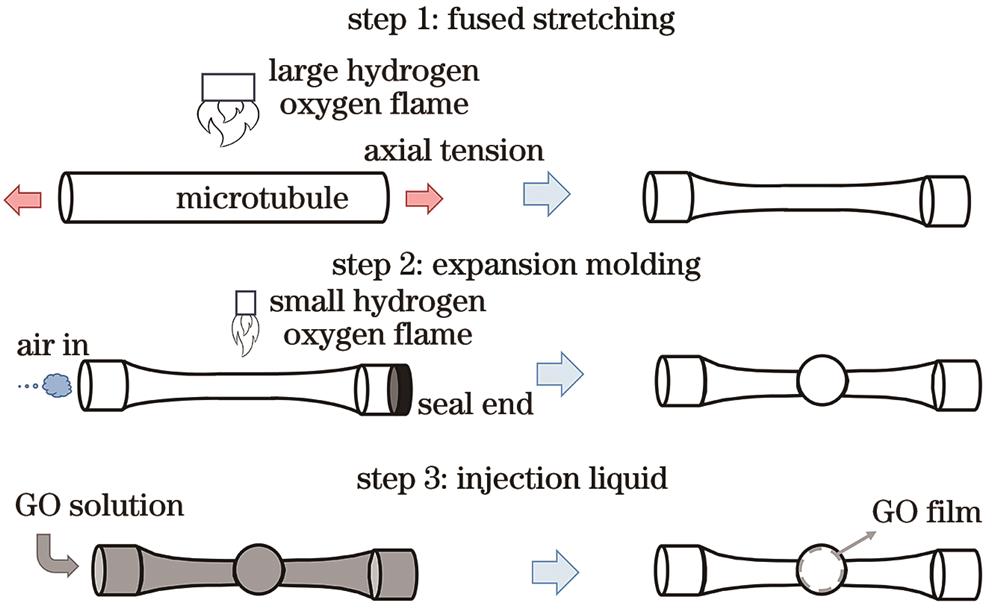
图 2. GO涂覆空心微泡腔气体传感器的制备步骤示意图
Fig. 2. Schematic diagram of fabrication steps for GO coated hollow microsphere cavity gas sensor
3.2 氨气传感系统搭建
氨气传感实验装置如
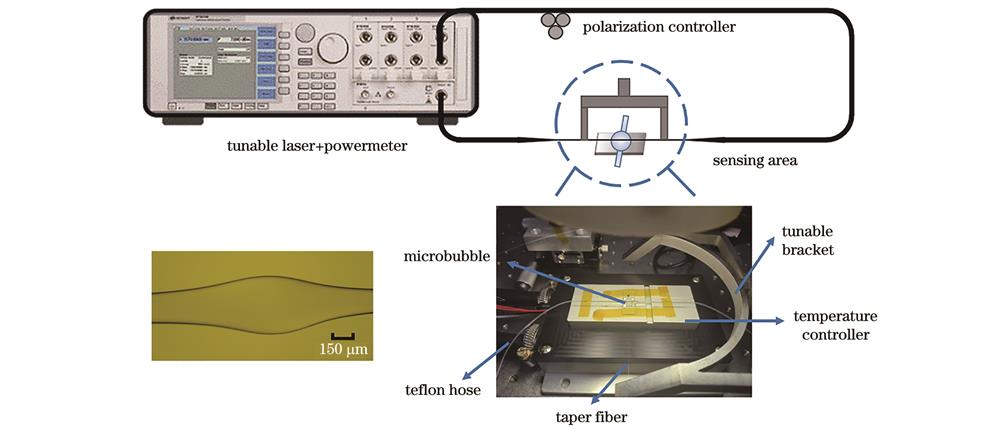
图 3. 基于GO涂覆微泡腔的气体传感实验装置示意图
Fig. 3. Schematic diagram of gas sensing experimental device based on GO coated microsphere cavity
4 结果分析与讨论
4.1 对氨气气体的响应以及灵敏度和分辨率结果
保持温控台的温度为25 ℃,向GO涂覆的空心微泡腔传感器内注入不同体积分数的NH3气体,体积分数在0~100×10-6之间,间隔为20×10-6。GO涂覆空心微泡腔的传感器WGM的透射谱随气体体积分数的移动如

图 4. GO涂覆微泡腔的WGM的光谱随气体体积分数漂移的实验测试结果。(a)不同体积分数的氨气对应的GO涂覆微泡腔的WGM的光谱;(b)中心波长漂移曲线
Fig. 4. Experimental test results of spectra of WGM with GO coated microsphere drifting with volume fraction. (a) Spectra of WGM with GO coated microsphere measured at different volume fraction of NH3; (b) center wavelength shift curves
为了进一步评估传感器的灵敏度和分辨率,选择中心波长变化近似线性的较小的体积分数范围进行测试,通入微泡腔的NH3体积分数为0~40×10-6,间隔为5×10⁻⁶。在每个气体体积分数下,待谐振峰稳定后测量10次中心波长,每次间隔30 s。输出光谱由功率计模块记录,中心波长由洛伦兹拟合得到。传感器的共振中心波长与NH3的体积分数变化关系如

图 5. 较小体积分数下GO涂覆微泡腔的WGM的光谱随气体体积分数漂移的实验测试结果。(a)不同体积分数的氨气对应的GO涂覆微泡腔的WGM传感器谐振峰的中心波长;(b)中心波长漂移曲线以及每个体积分数下的标准差
Fig. 5. Experimental test results of spectra of WGM with GO coated microsphere drifting with gas volume fraction at low volume fraction. (a) Center wavelength of WGM sensor resonance peak measured at different volume fraction of NH3; (b) drift curve of center wavelength and standard deviation for each volume fraction
此前的氨气气体传感实验中,微泡腔传感器被放置于高精度温控台上以避免温度波动带来的影响。而实际应用中,温度的波动不可避免。为了获得传感器的温度响应,控制高精度温控台的温度由20 ℃升高至30 ℃。每次温度改变1 ℃,等待1 min温度稳定后,采集WGM光谱。谐振峰中心波长随温度的变化如
4.2 氨气气体的响应和恢复时间实验
分别将体积分数为10×10-6、20×10-6和40×10-6的NH3与干燥空气周期性地注入微泡腔传感器中。
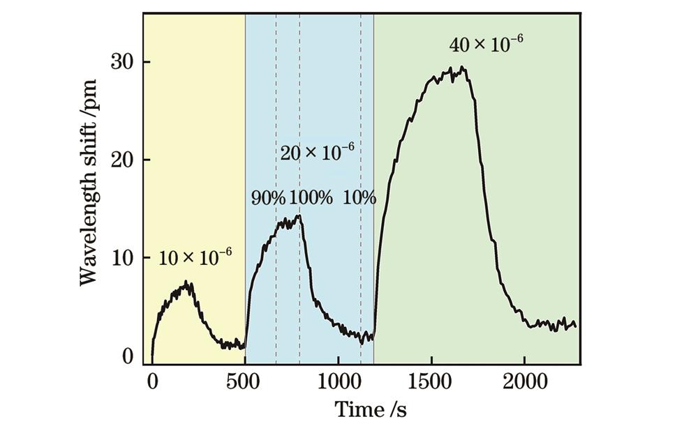
图 7. 对体积分数为10×10-6~40×10-6的NH3的实时响应曲线
Fig. 7. Real-time response curve for NH3 with volume fraction of 10×10-6-40×10-6
5 结论
本文设计了一种基于氧化石墨烯涂覆的空心微泡腔回音壁谐振模气体传感器,基于气体分子吸附对氧化石墨烯折射率的影响改变微腔整体结构的有效折射率,这反映为微腔所支持的回音壁谐振模漂移。通过功率计实时监控波长变化,以此进行气体传感。采用熔融加压流变成型及灌注氧化石墨烯分散液的方式制备实验所需的传感器。氨气气体传感实验结果表明,在低于40×10-6的气体体积分数内,传感器的灵敏度为0.73×10⁶ pm。结合整体系统的波长漂移标准差可以得到传感器的分辨率为1.9×10⁻⁶。在20×10-6气体体积分数下,传感器的响应和恢复时间分别是294 s和329 s。所提出的氧化石墨烯涂覆气体传感器同时具有高分辨率以及快速响应的优势。同时,空心的传感器结构使得其进行气体传感时无需额外的气体通道或气室封装结构,为实际应用提供了极大的便利。
[1] Novoselov K S, Geim A K, Morozov S V, et al. Electric field effect in atomically thin carbon films[J]. Science, 2004, 306(5696): 666-669.
[2] Bonaccorso F, Sun Z, Hasan T, et al. Graphene photonics and optoelectronics[J]. Nature Photonics, 2010, 4: 611-622.
[3] Fowler J D, Allen M J, Tung V C, et al. Practical chemical sensors from chemically derived graphene[J]. ACS Nano, 2009, 3(2): 301-306.
[4] Geim A K, Novoselov K S. The rise of graphene[J]. Nature Materials, 2007, 6: 183-191.
[5] Schedin F, Geim A K, Morozov S V, et al. Detection of individual gas molecules adsorbed on graphene[J]. Nature Materials, 2007, 6(9): 652-655.
[6] Yao B C, Wu Y, Cheng Y, et al. All-optical Mach-Zehnder interferometric NH3 gas sensor based on graphene/microfiber hybrid waveguide[J]. Sensors and Actuators B: Chemical, 2014, 194: 142-148.
[7] Rumyantsev S, Liu G X, Shur M S, et al. Selective gas sensing with a single pristine graphene transistor[J]. Nano Letters, 2012, 12(5): 2294-2298.
[8] 庾财斌. 基于氧化石墨烯的光纤干涉传感技术研究与应用[D]. 成都: 电子科技大学, 2018.
YuC B. Research and application of fiber-optic sensing technology based on graphene oxide[D]. Chengdu: University of Electronic Science and Technology of China, 2018.
[9] Compton O C, Nguyen S T. Graphene oxide, highly reduced graphene oxide, and graphene: versatile building blocks for carbon-based materials[J]. Small, 2010, 6(6): 711-723.
[10] Lerf A, He H Y, Forster M, et al. Structure of graphite oxide revisited[J]. The Journal of Physical Chemistry B, 1998, 102(23): 4477-4482.
[11] Chung M G, Kim D H, Lee H M, et al. Highly sensitive NO2 gas sensor based on ozone treated graphene[J]. Sensors and Actuators B: Chemical, 2012, 166/167: 172-176.
[12] Yasaei P, Kumar B, Hantehzadeh R, et al. Chemical sensing with switchable transport channels in graphene grain boundaries[J]. Nature Communications, 2014, 5: 4911.
[13] 李闯, 蔡理, 李伟伟, 等. 水合肼还原的氧化石墨烯吸附NO2的实验研究[J]. 物理学报, 2019, 68(11): 118102.
Li C, Cai L, Li W W, et al. Adsorption of NO2 by hydrazine hydrate-reduced graphene oxide[J]. Acta Physica Sinica, 2019, 68(11): 118102.
[14] 陈浩, 彭同江, 刘波, 等. 氧化石墨烯在气体传感器中的应用研究进展[J]. 材料导报, 2016, 30(17): 57-63.
Chen H, Peng T J, Liu B, et al. Research progress in gas sensors based on graphene oxide[J]. Materials Reports, 2016, 30(17): 57-63.
[15] Cittadini M, Bersani M, Perrozzi F, et al. Graphene oxide coupled with gold nanoparticles for localized surface plasmon resonance based gas sensor[J]. Carbon, 2014, 69: 452-459.
[16] Toda K, Furue R, Hayami S. Recent progress in applications of graphene oxide for gas sensing: a review[J]. Analytica Chimica Acta, 2015, 878: 43-53.
[17] Singh M, Raghuwanshi S K, Prakash O. Ultra-sensitive fiber optic gas sensor using graphene oxide coated long period gratings[J]. IEEE Photonics Technology Letters, 2019, 31(17): 1473-1476.
[18] 刘铁根, 于哲, 江俊峰, 等. 分立式与分布式光纤传感关键技术研究进展[J]. 物理学报, 2017, 66(7): 070705.
Liu T G, Yu Z, Jiang J F, et al. Advances of some critical technologies in discrete and distributed optical fiber sensing research[J]. Acta Physica Sinica, 2017, 66(7): 070705.
[19] Vahala K J. Optical microcavities[J]. Nature, 2003, 424(6950): 839-846.
[20] Foreman M R, Swaim J D, Vollmer F. Whispering gallery mode sensors[J]. Advances in Optics and Photonics, 2015, 7(2): 168-240.
[21] Liu Y Z, Jiang J F, Liu K, et al. Liquid-core-microtubule-enhanced laser sensor for high-resolution temperature measurement[J]. IEEE Sensors Journal, 2023, 23(13): 14167-14173.
[22] 张雪杨, 杨火木, 邓国亮, 等. 激光诱导石墨烯全光调谐超高Q值回音壁微腔[J]. 激光与光电子学进展, 2023, 60(23): 2314004.
[23] Gorodetsky M L, Savchenkov A A, Ilchenko V S. Ultimate Q of optical microsphere resonators[J]. Optics Letters, 1996, 21(7): 453-455.
[24] Gregor M, Pyrlik C, Henze R, et al. An alignment-free fiber-coupled microsphere resonator for gas sensing applications[J]. Applied Physics Letters, 2010, 96(23): 231102.
[25] Passaro V M N, Dell'Olio F, de Leonardis F. Ammonia optical sensing by microring resonators[J]. Sensors, 2007, 7(11): 2741-2749.
[26] Jiang X F, Qavi A J, Huang S H, et al. Whispering-gallery sensors[J]. Matter, 2020, 3(2): 371-392.
[27] Sun Y Z, Liu J, Frye-Mason G, et al. Optofluidic ring resonator sensors for rapid DNT vapor detection[J]. Analyst, 2009, 134(7): 1386-1391.
[28] Yao B C, Yu C B, Wu Y, et al. Graphene-enhanced Brillouin optomechanical microresonator for ultrasensitive gas detection[J]. Nano Letters, 2017, 17(8): 4996-5002.
[29] Paul R K, Badhulika S, Saucedo N M, et al. Graphene nanomesh as highly sensitive chemiresistor gas sensor[J]. Analytical Chemistry, 2012, 84(19): 8171-8178.
[30] Li Z Q, Henriksen E A, Jiang Z, et al. Dirac charge dynamics in graphene by infrared spectroscopy[J]. Nature Physics, 2008, 4: 532-535.
[31] Lee C Y, Strano M S. Understanding the dynamics of signal transduction for adsorption of gases and vapors on carbon nanotube sensors[J]. Langmuir, 2005, 21(11): 5192-5196.
[32] Peng Y, Li J H. Ammonia adsorption on graphene and graphene oxide: a first-principles study[J]. Frontiers of Environmental Science & Engineering, 2013, 7(3): 403-411.
[33] Vollmer F, Arnold S. Whispering-gallery-mode biosensing: label-free detection down to single molecules[J]. Nature Methods, 2008, 5(7): 591-596.
[34] Jiang J F, Liu Y Z, Liu K, et al. Wall-thickness-controlled microbubble fabrication for WGM-based application[J]. Applied Optics, 2020, 59(16): 5052-5057.
[35] Cai M, Painter O, Vahala K J. Observation of critical coupling in a fiber taper to a silica-microsphere whispering-gallery mode system[J]. Physical Review Letters, 2000, 85(1): 74-77.
[36] Yu Z, Wang S, Jiang J F, et al. Investigation of fused tapering with inner pressurized air for microcapillary-based optical sensor[J]. Optical Fiber Technology, 2018, 45: 244-249.
Article Outline
刘译泽, 江俊峰, 刘琨, 王双, 王逸璇, 陈鑫, 刘铁根. 氧化石墨烯涂覆的空心微泡腔回音壁谐振模气体传感器[J]. 光学学报, 2024, 44(7): 0728004. Yize Liu, Junfeng Jiang, Kun Liu, Shuang Wang, Yixuan Wang, Xin Chen, Tiegen Liu. Gas Sensor Based on Graphene Oxide-Coated Hollow Microbubble Whisper Gallery Resonant Mode[J]. Acta Optica Sinica, 2024, 44(7): 0728004.
