校正光通量变化的定量光声内窥成像  下载: 712次
Photoacoustic imaging (PAI) is an emerging imaging modality that provides structural and functional information on biological tissue, with the advantages of high contrast of optical imaging and high penetration depth of ultrasound imaging. Photoacoustic endoscopic imaging is an endoscopic application of PAI, which combines non-invasive PAI with endoscopic detection technology. This imaging modality is physically based on the photoacoustic effect of biological tissue illuminated by short laser pulses. The absorbed optical energy density is proportional to the product of the optical absorption coefficient and the local light fluence. Therefore, light fluence is related not only to the optical properties of the tissue but also to the distribution of irradiation intensity. The optical properties of the tissue cannot be accurately reflected in the distribution image of absorption energy density. By solving the optical inverse problem, the optical property parameters can be estimated quantitatively to realize quantitative imaging. Due to the complex optical properties of tissue and non-uniform and non-stationary illumination, photoacoustic image reconstruction is subject to non-uniform light fluence, which leads to a reduction in image quality and imaging depth. The purpose of this work is to solve the problem of reduced accuracy of quantitative imaging due to light fluence variation.
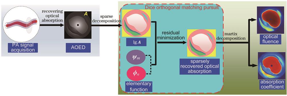
A quantitative image reconstruction method for photoacoustic endoscopic imaging is presented to correct light fluence variation. It employs a two-step scheme. Firstly, the distribution of absorbed optical energy density on the cross-section of the tubular object is recovered from the photoacoustic signal measured by the ultrasonic detector with conventional image reconstruction methods such as back projection and time reversion. Secondly, the sparse representation of the distribution of absorbed optical energy density is constructed by the weighted sum of a finite number of discrete Curvelet and Haar joint basis functions. The sparse representations of the absorption coefficient and light fluence are then obtained by the greedy algorithm. In addition, the absorption coefficient and light fluence distributions are reconstructed simultaneously by sparse matrix decomposition.
The method has been verified by simulation and phantom studies. The comparisons with the one-step method, model-based method, and state-of-the-art fluence compensation method demonstrate the superiority of the proposed method in recovering optical coefficient distribution with high accuracy. The simulations show that the one-step method has a large error when estimating the absorption coefficient, and the reconstructed image cannot distinguish different tissue types clearly. In the images reconstructed by the Bregman method, significant overlap between different tissue regions can be observed. The images reconstructed by the perturbation Monte Carlo method show the low contrast of tissue boundaries in the low-fluence region. The proposed method is superior to other methods in recovering the absorption coefficient closest to the ground truth. In the phantom study, due to similar optical properties of the two materials used to fabricate the phantom, the overall contrast of the image representing the distribution of absorbed optical energy density is low, and it is difficult to distinguish the contours of the embedded targets. In contrast, images showing the distribution of absorption coefficients can clearly distinguish the contours of the targets. In addition, compared with other methods, the proposed method can achieve the highest accuracy in absorption efficient estimation. Simulation and experimental results on the phantom reveal that the root mean square error (RMSE) of the absorption coefficient estimated with this method can be reduced by about 48%, and the normalized mean square absolute distance (NMSAD) and structural similarity (SSIM) of the reconstructed images can be reduced by about 25% and increased by about 24%, respectively, compared with the one-step method and model-based method. The comparisons with the state-of-the-art fluence compensation method show significant improvements in RMSE, NMSAD, and SSIM metrics by about 22%, 20%, and 10%, respectively.
This work presents a joint reconstruction method of the absorption coefficient and light fluence distribution based on sparse decomposition to construct the distribution of absorbed optical energy density. The method shows advantages over the one-step method based on exact solutions, the model-based method, and the state-of-the-art fluence compensation method in recovering absorption coefficient distribution with high accuracy. Furthermore, the quantitative reconstruction accuracy of the proposed method is not sensitive to the recovery of the absorbed optical energy density and the similarity measurement of sparse decomposition. It should be noted that the optimization algorithm for sparse reconstruction influences the reconstruction accuracy, among which the accuracy of multi-candidate orthogonal matching pursuit (MOMP) and Dice orthogonal matching pursuit (DOMP) is higher than that of matching pursuit (MP) and basis pursuit (BP). Future work should involve the following two aspects. One is to verify the feasibility of this method in clinical transplantation through in vivo experiments. The reconstruction accuracy should be further improved with comprehensive consideration of non-ideal factors in practical application scenarios, such as limited-view sparse-sampling measurement, acoustic reflection and scattering, motion artifacts caused by cardiac motion in intracoronary photoacoustic imaging, and characteristics of ultrasonic detectors. The other is to attempt to apply deep learning to eliminate the influence of light fluence variation on PAI quality.
孟琪, 孙正, 侯英飒, 孙美晨. 校正光通量变化的定量光声内窥成像[J]. 光学学报, 2023, 43(1): 0111001. Qi Meng, Zheng Sun, Yingsa Hou, Meichen Sun. Quantitative Photoacoustic Endoscopic Imaging for Correcting Light Fluence Variation[J]. Acta Optica Sinica, 2023, 43(1): 0111001.


