数字全息内窥技术实现耳膜病态下的振动模态研究
Ear is an important hearing organ of the human body. While hearing, the eardrum vibrates to transmit the incoming sound to the middle ear, and the characteristics of these vibrations have a direct impact on hearing. Studying the vibration parameters of the eardrum is extremely significant and valuable for the medical diagnosis of hearing disorders. In previous studies, researchers have used digital holographic technology to detect the amplitude and phase of the vibrating eardrum surface. However, because of the intricate location of the eardrum in the ear canal, the optical path structure is limited. Currently, the detection of key eardrum parameters relies primarily on dissected samples. The correlation between the eardrum defects and vibration modes obtained through experimental and simulation analyses remains unclear. Therefore, digital holographic endoscopy is proposed to study the vibration mode of a defective eardrum, and an algorithm for spatial carrier phase extraction is implemented to detect the vibration mode. Compared with the fringe pattern of the amplitude distribution obtained using the traditional image subtraction mode, the light and dark contrast of the fringes obtained by phase subtraction is significantly improved. In this study, the effects of different defects in an eardrum on the first-order vibration mode were verified, thereby providing a method and theoretical basis for the in?situ detection of defective eardrums.
In this study, the relationship between eardrum defects and vibration modes was analyzed using finite element simulation and experimental analysis. Owing to the difficulty in obtaining an eardrum, a silicone rubber film, which is commonly used in the medical eardrum repair, was used as a substitute. In the finite element analysis, we studied the changes in the first-order vibration mode of the artificial eardrum based on perforation, scratch and calcification. In the vibration mode detection experiment, we built an optical path of the Mach-Zehnder interferometer for digital holographic endoscopy and used a sinusoidal signal excited by a speaker to stimulate the resonance of the sample surface. Spatial carrier phase extraction based on the time-averaged method was used to detect the amplitude distribution of the film surface in the vibration mode. Based on the changes observed in the amplitude distribution in the first-order vibration mode for the artificial eardrums with different defects, the location and severity of the defect and their influence on the vibration were analyzed.
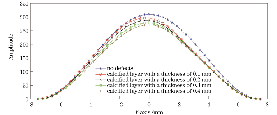
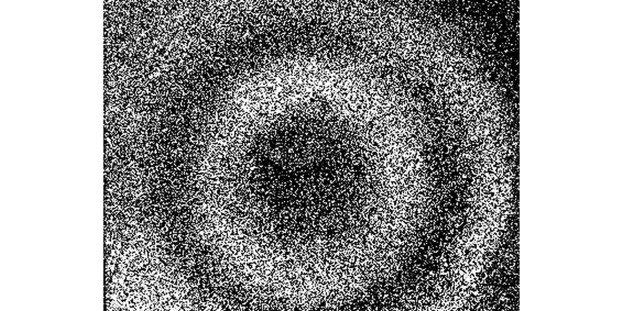
First, the theoretical analysis proves that using the spatial carrier phase extraction method to detect the amplitude distribution in eardrum samples in the vibration mode is reasonable. In the finite element simulation and experimental analysis, the vibration modes of the artificial eardrums with defects were analyzed, and the results showed that different defects affect the amplitude distribution in the first-order vibration mode for the eardrums differently. For the perforated eardrum samples, the amplitude distribution was analyzed by varying the size and location of the perforation. The results show that the amplitude near the perforation increases significantly with the increase in perforation size (Figs. 3 and 4), and an increase in the number of fringes is observed in the experimental results (Fig. 11). By changing the location of the perforation, the maximum amplitude shifts off-center with the perforation, and the larger the perforation, the more evident the deviation. The amplitude distribution for the scratched eardrum samples was analyzed by varying the size and location of the scratch. The results show that at the same location, the larger the scratch length, the larger the surface amplitude of the film (Fig. 5), and the amplitude changes more significantly near the scratch location (Fig. 6). The experimental results show an increase in the number of fringes, and the shape of the fringe near the central scratch is flat (Fig. 12). When the scratch is off-center, the effect on the amplitude near the scratch is significantly greater than that at the center. The amplitude distribution for the calcified eardrum samples was analyzed by varying the thickness of the calcified layer (Fig. 7). The amplitude of the film decreases with an increase in the thickness of the calcified layer but is more evident at the location of the calcified layer (Figs. 8 and 13).
In this study, a finite element simulation method was used to evaluate the influence of different defects of an eardrum on the first-order vibration mode. Digital holographic endoscopy was used to detect the vibration mode of the eardrum, and experimental verification of the simulation results was performed. The simulations and experimental results show that variations in the defects of the eardrum affect the first-order vibration mode, and the effects differ based on the size and location of the defect. From a mechanical perspective, an eardrum defect leads to a change in the local stiffness of the structure. Perforation and scratches reduce the stiffness, and calcification increases the stiffness; this leads to increased vibration of the eardrum near the perforated and scratched regions and a weakened vibration near the calcification layer. This study shows the influence of defects on the vibration of the eardrums by analyzing the distribution of the amplitude at the eardrum and the location and severity of the defect. This study provides an optical detection technique for evaluating eardrum defects, which can help in preventing and detecting hearing disorders.
1 引言
耳朵是人体重要的听觉器官,耳膜在整个听觉系统中的主要作用是将接收到的外界声压信号转换为耳膜的振动信号,最终产生听觉[1]。在整个过程中,耳膜的振动反映了中耳的传声性能,其振动性能对人的听力具有直接影响,耳膜对振动的响应可表现为耳膜表面形变、振型分布、振动频率的变化。耳膜结构复杂,且位于耳道内部,其产生的表面形变又通常在微米量级,因此需要借助有效的技术手段来检测耳膜的相关参数。
在此之前,相关学者已经对耳膜进行了广泛研究,主要有实验检测和有限元仿真分析。在实验检测方面,人们采用的方法主要有激光多普勒测振技术、数字图像相关技术、时间平均或频闪全息测量技术、光学相干断层扫描法等,这些技术为检测耳膜振动机制提供了重要工具[2]。1987年,Konrádsson等[3]使用扫描激光多普勒测振仪记录并重建了单频率下具有振幅和相位图的人类耳膜三维振动。2002年,Wada等[4]将正弦相位调制应用于时间平均散斑干涉测量中,以中等速度检测了豚鼠耳膜整个表面的振幅和相位。2018年,Gladiné等[5]先使用调色剂和荧光粉在耳膜上创建斑点图案并进行染色,然后利用数字图像相关技术进行重建,实现了完整耳膜的形变和应变测量。2019年,Psota等[6]在高速全息系统的基础上提出了一种新的照明技术,并基于该技术利用单波长高功率激光器对耳膜进行了形状测量。应用上述方法可以检测耳膜振动信号,并能够辅助研究中耳出现病变时造成的听力损失。在有限元仿真分析方面,人们通常通过对中耳的几何参数和力学性能进行模拟分析来计算耳膜在声压下的振动情况。2007年,Gan等[7]利用有限元分析了鼓室内的流体对鼓膜和镫骨底板的位移以及中耳传声的影响。2004年,Gan等[8]根据建立的有限元模型,分析了耳膜弹性模量和厚度对其振动性能的影响。2015年,王杰等[9]针对不同厚度耳膜对中耳传声的影响进行了有限元分析,结果发现耳膜厚度会影响中耳的传声效能。
综上,由于耳膜在耳道内的位置比较特殊,对光路结构具有一定的限制性,目前耳膜关键参数的检测主要是针对离体解剖样本进行的,而且现有的实验和仿真分析都未明确耳膜缺陷和耳膜振动模态之间的关联性。
由于物体在共振下的各阶振型是恒定的,其缺陷会引起振型的变化,而数字全息技术具有无损、全场和动态测量等优势,在形变检测、生物医学成像等方面被广泛应用[10-12],因此,笔者提出了用于研究耳膜病态下振动模态的数字全息内窥技术,并将空间载波相位提取方法应用到耳膜的振动模态检测中,为实现耳膜病态的原位检测提供了实现途径和理论依据。为简化实验条件,笔者针对外伤性耳膜病态主要发生在紧张部这一实际情况[13],将人造耳膜材料硅橡胶薄膜作为实验样本,就耳膜的穿孔、划伤和钙化缺陷对一阶振动模态的影响进行研究;然后通过有限元仿真分析了不同病态耳膜一阶振型和振幅分布的变化,并采用数字全息技术获取了一阶振动模态下的振幅分布相位条纹图;最后采用实验验证了仿真方法的有效性。
2 薄膜振动模态检测原理
2.1 薄膜自由振动原理
假设圆形薄膜厚度均匀,半径为
根据哈密顿原理,在极坐标
式中:
薄膜振动的边界条件为
先后令
式中:
式中:k0为常系数。
根据
式中:
设
式中:
由角频率
2.2 离面形变数字全息检测原理
采用马赫-曾德尔数字全息内窥干涉光路实现耳膜离面形变信息的全息图记录。
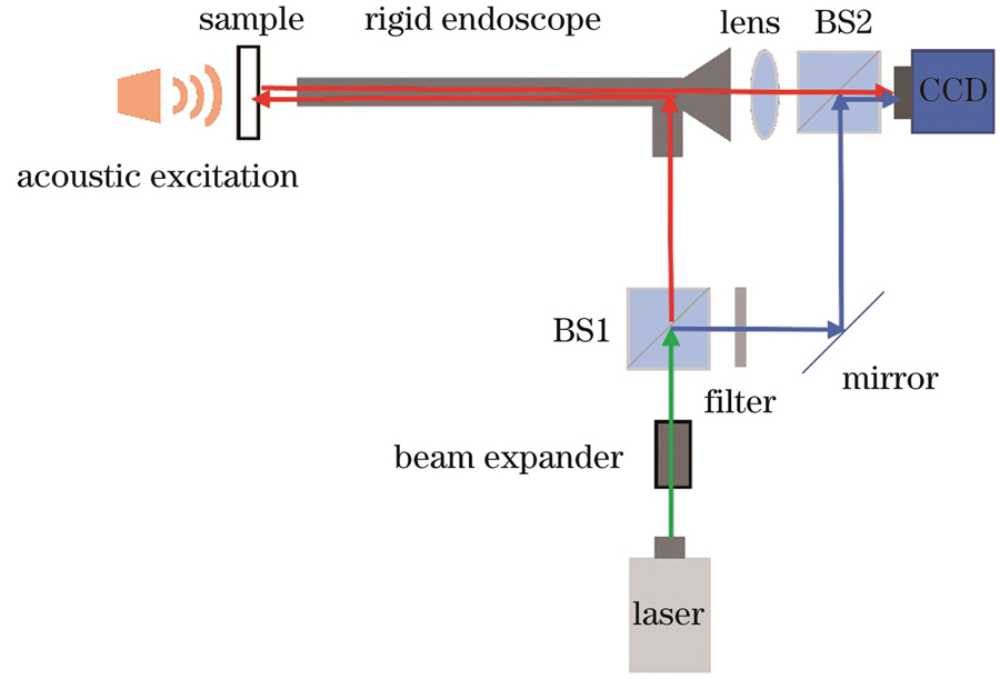
图 2. 数字全息内窥干涉光路示意图
Fig. 2. Schematic of digital holographic endoscopy interference light path
物体在静止状态时,CCD记录的强度图表达式为
式中:
当物体作纯离面振动时,振动相位可以表示为
式中:
根据时间平均原理,当CCD曝光时间为振动周期的整数倍时,物体振动后的强度图表达式为
其中,
根据
为获取模态下的振幅分布信息,将
由于相位表达式中的第一类零阶贝塞尔函数只有0和
3 仿真及实验
3.1 病态对人造耳膜材料薄膜振动模态影响的仿真分析
真实耳膜的采集较为困难,因此笔者采用医学耳膜修复中常用的人造耳膜材料硅橡胶薄膜作为代替品,采用有限元仿真软件分析了穿孔、划伤和钙化情况下人造耳膜材料薄膜一阶振动模态的变化。真实耳膜近似为圆形半透膜,直径为8~9 mm,厚度约为0.1 mm。相关研究表明,圆形薄膜在振动模态下的振动位移与其直径、厚度呈线性关系[15,17]。因此,为便于实验样本的固定,利用人造耳膜材料制作了直径为15 mm、厚度为0.3 mm的圆形薄膜作为耳膜样本,其钙化缺陷采用钙化层材料制成。人造耳膜和钙化层的材料参数如
表 1. 材料属性
Table 1. Material attribute
|
通过改变穿孔和划伤的大小、位置以及钙化层厚度分析了耳膜病态对一阶振动模态的影响。需要说明的是,仿真软件中给出的振动位移不具备单位级别,数据之间仅为相对大小关系。
在薄膜上设置圆形缺陷来模拟耳膜穿孔。
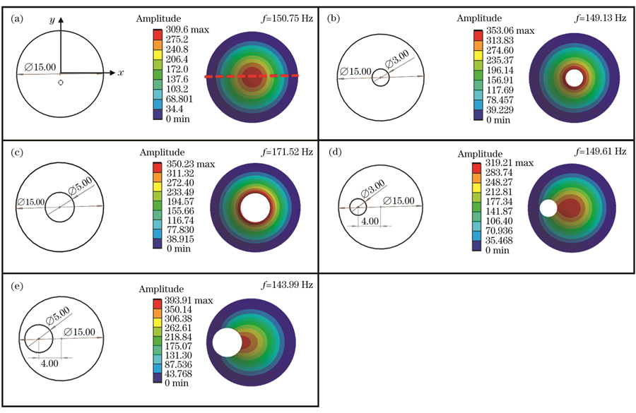
图 3. 穿孔薄膜一阶振动模态的仿真结果。(a)无缺陷薄膜;(b)中心穿孔3 mm薄膜;(c)中心穿孔5 mm薄膜;(d)偏心穿孔3 mm薄膜;(e)偏心穿孔5 mm薄膜
Fig. 3. Simulation results of first-order vibration modes of perforated films. (a) Defect-free film; (b) film with a 3 mm diameter perforation in the center; (c) film with a 5 mm diameter perforation in the center; (d) film with an eccentric 3 mm diameter perforation; (e) film with an eccentric 5 mm diameter perforation
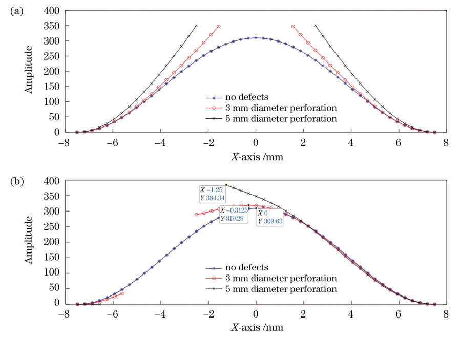
图 4. y=0处的振幅分布截面图。(a)中心穿孔薄膜;(b)偏心穿孔薄膜
Fig. 4. Section of amplitude at y=0. (a) Central perforated film; (b) eccentric perforated film
如
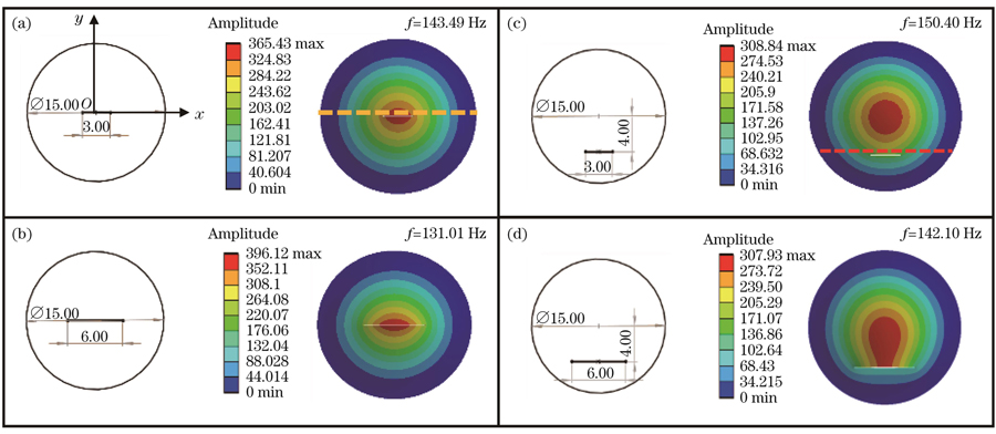
图 5. 划伤薄膜一阶振动模态的仿真结果。(a)中心3 mm划伤薄膜;(b)中心6 mm划伤薄膜;(c)偏心3 mm划伤薄膜;(d)偏心6 mm划伤薄膜
Fig. 5. Simulation results of first-order vibration modes of scratched films. (a) Film with a 3 mm scratch in the center; (b) film with a 6 mm scratch in the center; (c) film with an eccentric 3 mm scratch; (d) film with an eccentric 6 mm scratch
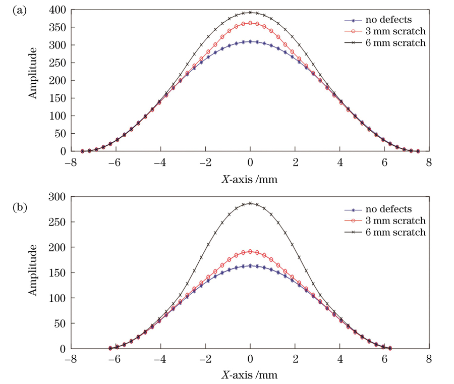
图 6. 划伤薄膜振幅分布截面图。(a)中心划伤薄膜y=0.1处;(b)偏心划伤薄膜y=-3.7处
Fig. 6. Section of amplitude of scratched films. (a) Section at y=0.1 of the centrally scratched film; (b) section at y=-3.7 of the eccentrically scratched film
耳膜钙化常常是由耳部炎症引起的,表现为耳膜表面白色斑块状钙盐沉积,钙化严重的情况下会导致听力下降。如
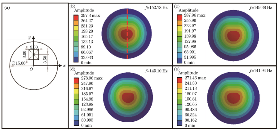
图 7. 钙化薄膜一阶振动模态的仿真结果。(a)钙化薄膜示意图;(b)~(e)钙化厚度为0.1、0.2、0.3、0.4 mm的薄膜
Fig. 7. Simulation results of first-order vibration modes of calcified films. (a) Schematic of calcified film; (b)-(e) films with calcification thickness of 0.1, 0.2, 0.3 and 0.4 mm, respectively
3.2 病态对人造耳膜材料薄膜振动模态影响的实验验证
根据
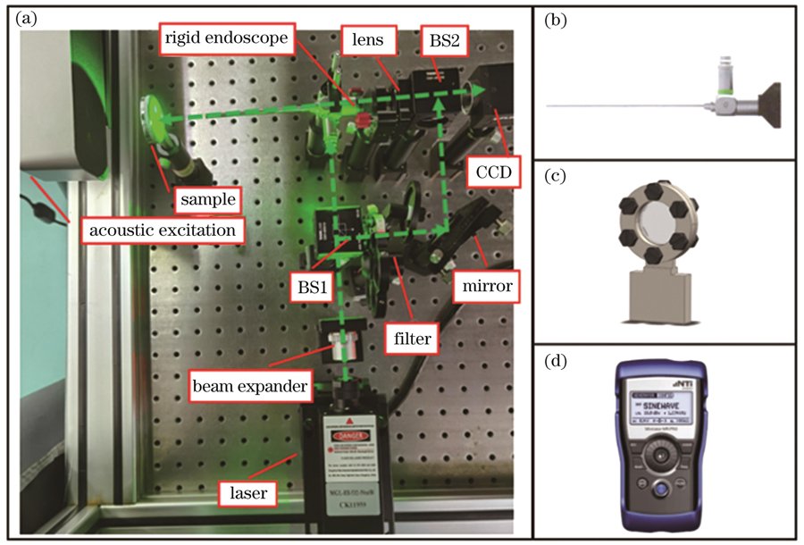
图 9. 离面形变检测实验装置。(a)光路系统;(b)J0900D内窥耳镜;(c)样本夹具;(d)音频信号发生器
Fig. 9. Experimental device for out-of-plane deformation detection. (a) Optical path system; (b) J0900D endoscopic otoscope; (c) sample fixture; (d) sound generator
根据上述仿真,用
实验样本与仿真时的样本保持一致,
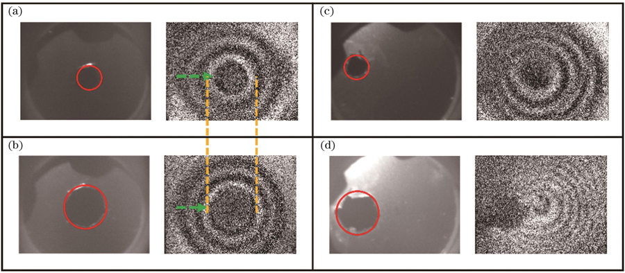
图 11. 穿孔薄膜的一阶振动模态图。(a)中心穿孔3 mm薄膜;(b)中心穿孔5 mm薄膜;(c)偏心穿孔3 mm薄膜;(d)偏心穿孔5 mm薄膜
Fig. 11. First-order vibrational mode diagrams of perforated films. (a) Film with a 3 mm diameter perforation in the center; (b) film with a 5 mm diameter perforation in the center; (c) film with an eccentric 3 mm diameter perforation; (d) film with an eccentric 5 mm diameter perforation
针对上述的薄膜划伤仿真分析,同样进行了相应的实验验证,得到的中心划痕样本的一阶振动模态图如
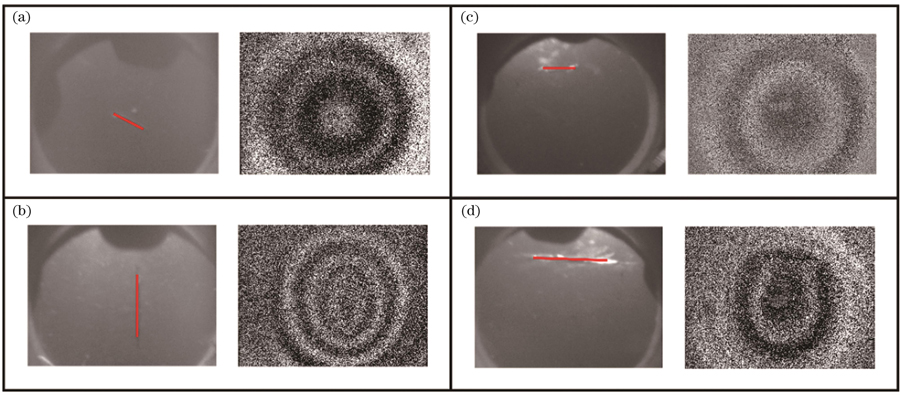
图 12. 划伤薄膜的一阶振动模态图。(a)中心3 mm划伤薄膜;(b)中心6 mm划伤薄膜;(c)偏心3 mm划伤薄膜;(d)偏心6 mm划伤薄膜
Fig. 12. First-order vibrational mode diagrams of scratched films. (a) Film with a 3 mm scratch in the center; (b) film with a 6 mm scratch in the center; (c) film with an eccentric 3 mm scratch; (d) film with an eccentric 6 mm scratch
在钙化薄膜的实验验证中,为了模拟真实的耳膜钙化情况,在薄膜表面涂抹不同厚度的尺寸为3 mm×3 mm的钙化材料。分别在相同位置处涂抹0.1 mm和0.3 mm厚度的钙化材料,如

图 13. 钙化薄膜的一阶振动模态图。(a)钙化厚度为0.1 mm的薄膜;(b)钙化厚度为0.3 mm的薄膜
Fig. 13. First-order vibrational mode diagrams of calcified films. (a) Film with calcification thickness of 0.1 mm; (b) film with calcification thickness of 0.3 mm
4 结论
为了方便研究耳膜病态对耳膜振动模态的影响,笔者选择常用的人造耳膜材料硅橡胶替代耳膜材料,通过有限元仿真软件研究了耳膜病态对耳膜一阶振动模态的影响,并采用数字全息内窥技术对仿真结果进行了相应的实验验证。仿真分析和实验研究结果表明,耳膜病态会对耳膜的一阶模态产生影响,且病态的尺寸和位置不同,产生的影响也不同。从力学角度来说,耳膜病态会导致其结构局部的刚度发生变化,穿孔和划伤会使刚度降低,钙化则会使刚度增大,进而导致薄膜在穿孔和划伤附近振动加剧,而在钙化层附近振动减弱。本研究说明病态会对耳膜振动产生影响,从而导致耳膜的振动性能发生改变,这势必会导致听力受损。本研究为耳膜病态研究提供了一种光学无损原位检测手段,对于听力方面疾病的预防和检测具有一定的参考价值。
[1] 王立坚, 王杰, 张李芳, 等. 两种声刺激模式下的正常鼓膜振动特征初步研究[J]. 中国听力语言康复科学杂志, 2019, 17(1): 13-16.
Wang L J, Wang J, Zhang L F, et al. A pilot study on the characteristics of normal tympanic membrane vibration in two types of acoustic stimulation modes[J]. Chinese Scientific Journal of Hearing and Speech Rehabilitation, 2019, 17(1): 13-16.
[2] 张李芳, 王杰, 李永新. 鼓膜振动研究技术进展[J]. 国际耳鼻咽喉头颈外科杂志, 2019, 43(3): 130-133.
Zhang L F, Wang J, Li Y X. Advances in research on tympanic membrane vibration[J]. International Journal of Dermatology and Venereology, 2019, 43(3): 130-133.
[3] Konrádsson K S, Ivarsson A, Bank G. Computerized laser Doppler interferometric scanning of the vibrating tympanic membrane[J]. Scandinavian Audiology, 1987, 16(3): 159-166.
[4] Wada H, Ando M, Takeuchi M, et al. Vibration measurement of the tympanic membrane of Guinea pig temporal bones using time-averaged speckle pattern interferometry[J]. The Journal of the Acoustical Society of America, 2002, 111(5): 2189-2199.
[5] Gladiné K, Dirckx J J J. 3D deformation and strain measurement of an intact eardrum using digital image correlation[J]. Journal of Physics: Conference Series, 2018, 1149(1): 012026.
[6] Psota P, Tang H M, Pooladvand K, et al. Investigation of tympanic membrane shape using digital holography[J]. Proceedings of SPIE, 2019, 11385: 113850G.
[7] Gan R Z, Wang X L. Multifield coupled finite element analysis for sound transmission in otitis media with effusion[J]. The Journal of the Acoustical Society of America, 2007, 122(6): 3527-3538.
[8] Gan R Z, Feng B, Sun Q L. Three-dimensional finite element modeling of human ear for sound transmission[J]. Annals of Biomedical Engineering, 2004, 32(6): 847-859.
[9] 王杰, Zhao Fei, 李永新. 颞肌筋膜重建鼓膜厚度对中耳传声的影响: 有限元模型研究[J]. 中国耳鼻咽喉头颈外科, 2015, 22(8): 414-418.
Wang J, Zhao F, Li Y X. Effect of tympanic membrane thickness in fascia myringoplasty on the middle ear transfer function: a finite element ear model[J]. Chinese Archives of Otolaryngology-Head and Neck Surgery, 2015, 22(8): 414-418.
[10] 满天龙, 万玉红, 菅孟静, 等. 面向生物样品三维成像的光干涉显微技术研究进展[J]. 中国激光, 2022, 49(15): 1507202.
[11] 张美娟, 夏海廷, 宋庆和, 等. 多相机数字全息测量物体三维变形方法研究[J]. 激光与光电子学进展, 2022, 59(16): 1609001.
[12] 刘雅坤, 肖文, 车蕾平, 等. 基于数字全息显微层析的癌细胞空泡化成像研究[J]. 中国激光, 2022, 49(20): 2007209.
[13] 包永新. 外伤性鼓膜穿孔临床治疗的研究[J]. 中国临床实用医学, 2007, 1(7): 41-42.
Bao Y X. Study of the clinical treatment of traumatic tympanic membrane perforation[J]. China Clinical Practical Medicine, 2007, 1(7): 41-42.
[14] 王靖义. 薄膜结构基于附加质量的振动特性分析[D]. 哈尔滨: 哈尔滨工业大学, 2021: 1-57.
WangJ Y. Vibration characteristics analysis of membrane structure based on additional mass[D]. Harbin: Harbin Institute of Technology, 2021: 1-57.
[15] 耿臻岑. 带缺陷蜂窝夹层结构的力学性能实验研究[D]. 南京: 东南大学, 2016: 1-63.
GengZ C. Experimental study on mechanical properties of honeycomb sandwich structure with defects[D]. Nanjing: Southeast University, 2016: 1-63.
[16] Picart P, Leval J, Mounier D, et al. Some opportunities for vibration analysis with time averaging in digital Fresnel holography[J]. Applied Optics, 2005, 44(3): 337-343.
[17] 谢传喜. 圆形薄膜结构在冲击荷载作用下的动力响应研究[D]. 重庆: 重庆大学, 2014: 1-70.
XieC X. Study on dynamic response of circular membrane structure under impact load[D]. Chongqing: Chongqing University, 2014: 1-70.
Article Outline
丁剑雯, 周文静, 于瀛洁. 数字全息内窥技术实现耳膜病态下的振动模态研究[J]. 中国激光, 2023, 50(15): 1507204. Jianwen Ding, Wenjing Zhou, Yingjie Yu. Vibration Modes Study of Defective Eardrum Realized Using Digital Holographic Endoscopy[J]. Chinese Journal of Lasers, 2023, 50(15): 1507204.
