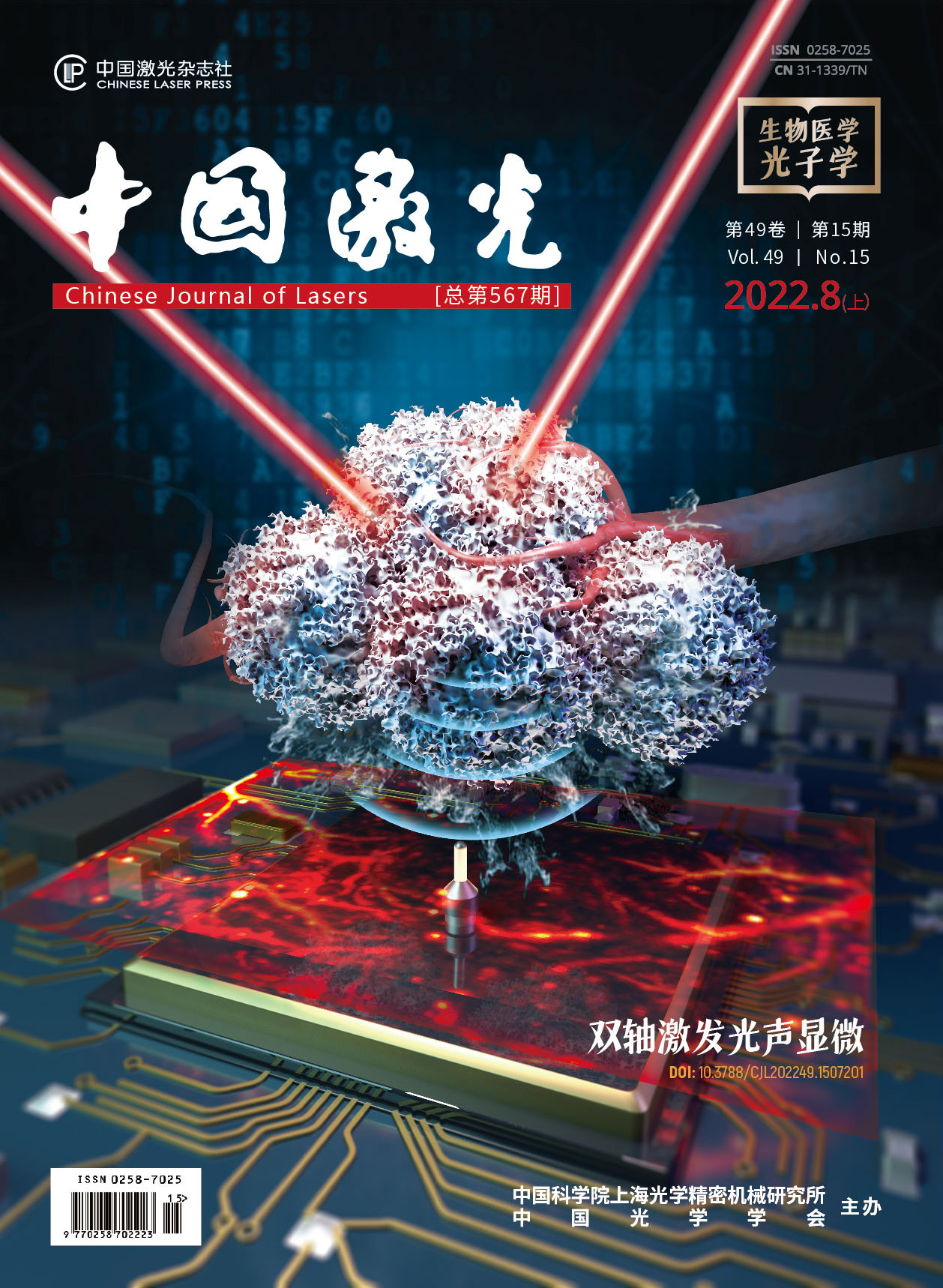
鲜红斑痣病灶结构及其光学成像方法在血管靶向光动力治疗中的研究进展  下载: 917次特邀综述亮点文章
Port wine stain (PWS), a skin disease with an incidence of 0.3%-0.5% in infants, is attributed to congenital and progressive cutaneous vascular malformations. PWS lesions appear as purplish red or pink patches on skin. PWS commonly occurs on the face and neck, but it can also appear anywhere on the body (e.g., arms or legs). PWS lesions give patients an extremely severe psychological burden about their cosmetic appearance, which may greatly reduce their life quality. The majority of PWS lesions darkens in color and thickens as time goes by. PWS lesions can become aggravated, leading to progressive disfigurement. Thus, early intervention and treatments are urgent to reduce the likelihood and severity of disfigurement and psychosocial morbidity.
The key point of treating PWS is to selectively and precisely destroy the abnormal blood vessels that cause the discolored lesions. Vascular-targeted photodynamic therapy (V-PDT) has been introduced and remarked as the golden standard of the therapy for PWS in China since the early 1990s, which destroys the ectatic vessesls by using the double selectivity of light and photosensitizer. Several clinical studies show that V-PDT is safe and effective in the treatment of PWS at all ages. Nevertheless, there are still some patients who have relatively low sensitivity to V-PDT. The unsatisfied therapeutic outcome may result from the heterogeneity and complexity of the PWS morphological parameters. Therefore, mastering the relationship between PWS lesion structure and V-PDT efficacy is beneficial to the development of therapeutic plan and the assessment of prognosis.
Skin biopsy is considered as the golden standard of acquiring the information of PWS lesion structure. Nevertheless, it is invasive which may lead to hypersensitivity to local anaesthetic agents, pain of local anaesthesia, bleeding, infection, and scarring. Hence, a painless and non-invasive imaging method for the in vivo assessment of PWS lesion structure will greatly benefit V-PDT treatments. With the development of optical imaging techniques, nowadays, they have showed much potential in obtaining the information of histopathology. Here, the relationship between PWS lesion structure (epidermis thickness, vascular diameter and depth, etc.) and V-PDT efficacy is summarized. In addition, the development of optical imaging techniques for obtaining PWS lesion structure is also reported.
Optical coherence tomography (OCT) called 'optical biopsy’ has been greatly developed in recent 20 years. Conventional OCT is used to visualize the fine structures of skin tissue although it is not an ideal tool for visualizing cutaneous vessels. As a functional modality of OCT, optical Doppler tomography (ODT) and optical coherence tomography angiography (OCTA) with the use of conjunction with sophistic algorism can provide a powerful tool to visualize and quantitatively analyze the cutaneous vascular networks. ODT and OCTA enhance the blood flow contrast by extracting moving particles (red blood cells) from static tissues without dye injection. To sum up, OCT/ODT/OCTA are capable of acquiring the epidermis thickness, the diameter and depth of vessels. Moreover, ODT/OCTA can further image a three-dimensional (3D) vascular system. However, OCT/ODT/OCTA have a relatively shallow imaging depth (less than 1 mm) because it is based on the optical imaging mechanism.
Photoacoustic imaging (PAI) combines the optical and ultrasonic merits is used to reduce the tissue scattering of photons with one-way ultrasound detection, while retaining the high optical contrast. Moreover, it can image over a 1 mm depth in skin. As an optical-contrast-based imaging technique, the photoacoustic imaging detects the endogenous skin chromophores, i.e., melanin and haemoglobin. PAI can obtain the content of melanin in epidermis, epidermis thickness, and vascular diameter and depth. Moreover, it can also achieve a 3D image of PWS vessels. However, PAI is a contact technique, in which a coupling medium (such as water and ultrasound gel) is required between the detector and tissue surface. Thus, it is unsuitable for an intraoperative use.
Reflectanceconfocal microscopy (RCM) is also a commonly used clinical skin diagnostic device. It uses a low-intensity near-infrared laser to first scan the skin horizontally, and then collects the backscattered light for imaging. RCM is the relatively mature imaging equipment and it can obtain the depth and diameter of skin vessels, nevertheless, it has the drawbacks of two-dimensional (2D) imaging and shallow imaging depth (200-350 μm).
Dermoscope is an imaging device that can magnify tens or even hundreds of times and eliminate the reflected light on the skin surface. Skin pigment and blood vessels are the two main elements of its observation, which have been widely used in the clinical practice. Although the dermoscope is convenient to use and fast to image, its imaging depth is limited (~250 μm), which cannot completely reflect the overall structure of PWS lesions, and the PWS manifestations under the dermoscope are irregular. Currently, the clinical application of dermoscope in PWS is still few.
Laser Doppler imaging (LDI) and laser speckle imaging (LSI) are able to get the information of blood perfusion, however, they only offer 2D information and the resolution is relatively low.
By applying the above techniques to obtain PWS lesion structure or biopsy, the relationship between PWS lesion structure and V-PDT efficacy is obtained as follows. In terms of melanin content in epidermis, it has the negative effect towards the V-PDT efficacy. It locates at the epidermis, absorbs the light, and influences optical absorbance. As for the epidermis thickness, it varies for different individuals and parts of the body, and affects the light penetration depth. Hence, it also has the negative effects towards V-PDT efficacy. In terms of vessels diameter, the larger the vessels, the less light can reach and cover the entire vessel. As for the vascular depth, the deeper the vessels are, the less light energy the vessels can absorb. The vascular morphological parameters also influence the efficacy. If the ratio of perpendicular vessels is higher than that of the curved ones, the efficacy is reduced due to the fact that the perpendicular vessels absorb less light than the curved ones. As for the blood perfusion, it is the mix-information which can be detected by LDI and LSI, and it may contain the information of diameter and depth. It is reported that if it decreases after V-PDT, the efficacy will be decent.
This paper summarizes the current application of the non-invasive in vivo optical imaging technology in the diagnosis and treatment of PWS as well as the relationship between PWS lesion structure and V-PDT efficacy. By mastering the above information, the clinical doctors can develop an accurate and personalized V-PDT treatment of PWS and thus improve the therapeutic effects.
刘一荻, 陈德福, 曾晶, 邱海霞, 顾瑛. 鲜红斑痣病灶结构及其光学成像方法在血管靶向光动力治疗中的研究进展[J]. 中国激光, 2022, 49(15): 1507102. Yidi Liu, Defu Chen, Jing Zeng, Haixia Qiu, Ying Gu. Progress in Port-Wine Stains Lesion Structure and Its Optical Imaging Technique in Vascular-Targeted Photodynamic Therapy[J]. Chinese Journal of Lasers, 2022, 49(15): 1507102.


