Sm3+掺杂NaYF4和BiPO4单颗粒微米晶体发光偏振特性研究  下载: 544次特邀研究论文
1 引言
由于稀土离子具有独特的4f壳层内部电子跃迁特性,以微纳米晶体为固态掺杂基质的稀土发光体被广泛地应用于各类微纳光学器件和光量子学的研究中[1-10]。虽然目前已有大量的研究报道涉及稀土微纳米晶体发光,但主要是基于其系综(粉末、胶体、溶液)发光特性的研究和应用[11-15]。近年来,得益于超分辨成像技术和弱光探测技术的快速发展,单颗粒光谱技术被用于检测单个稀土微纳米晶体的发光特性[16]。相较于常规系综光谱的平均效应,单颗粒光谱能够分辨单个发射体的特异性荧光指纹。利用该技术,研究人员发现稀土单颗粒晶体具有偏振发光特性[17-32],其与晶体的空间取向直接相关,这在系综光谱中常被掩盖和忽略。
偏振发光特性是稀土晶体唯一的矢量光学特性。早期,研究人员通过对比稀土离子同一光学跃迁,沿块状单轴单晶不同方向的偏振发射强度,判断光学跃迁的电磁振荡类型[33-40]。此外,通过检测稀土离子不同光学跃迁的偏振方向,可辅助检验离子所处的晶体场(点群)对称性[41]。截至目前,虽然有数十篇文献报道了单颗粒微纳米晶体中稀土离子的偏振发光现象,讨论和展望了其在单颗粒三维取向实时传感[29]、超晶格重构原位监测[30]、微流体中局部剪切速率实时测量等一系列前沿技术中的优势与潜力[31-32]。但是,这些工作主要集中在单一六方相晶体偏振发光特性的研究和应用开发上,尚缺乏对不同结构稀土单颗粒晶体偏振发光特性的研究和比较。
本文利用高精度单颗粒光谱表征技术,结合两种偏振检测方法,研究稀土铕离子掺杂单个六方相和单斜相结构晶体的偏振发光特性。实验发现,两种晶相的稀土单颗粒均发射部分偏振光,且所有发射峰的线偏角取决于单颗粒的取向。基于晶体宏观对称性和掺杂稀土离子的点群对称性,分析、阐明了这些现象的物理机制。
2 实验
2.1 实验原料
实验用药品包括Bi(NO3)3∙5H2O(纯度为99.5%)、NH4H2PO4(纯度为99.5%)、Sm(NO3)3∙6H2O(纯度为99.5%)、NaF(纯度为99.5%)、Y(NO3)3∙6H2O(纯度为99.5%)、乙二胺四乙酸(EDTA,纯度为99.5%)、无水乙醇(分析纯)、稀硝酸、去离子水,均购自国药集团化学试剂有限公司(上海),使用过程中均未经进一步提纯。
2.2 微米晶体制备
本实验采用水热法制备六方棒状NaYF4∶5%Sm3+微米晶体[42]。具体过程如下:首先,将1.365 g Y(NO3)3∙6H2O和0.083 g Sm(NO3)3∙6H2O溶解在15 mL去离子水中,制成稀土硝酸盐混合溶液。然后,将0.698 g EDTA和1.890 g NaF分别溶于20 mL和40 mL去离子水中,并依次将这两种溶液逐滴加入至上述稀土硝酸盐混合溶液中,利用磁力搅拌器搅拌混合溶液1 h。待搅拌完成,将其转移至带有聚四氟乙烯内衬的不锈钢反应釜中,在干燥箱中180 ℃加热24 h。待反应釜自然冷却至室温,利用去离子水和无水乙醇离心洗涤反应产物,可得六方棒状NaYF4∶Sm3+微米晶体粉末。
采用相似的方法制备单斜相BiPO4∶5%Sm3+微米晶体[43]。具体流程如下:首先,将1.728 g Bi(NO3)3∙5H2O和0.083 g Sm(NO3)3∙6H2O混入35 mL去离子水中,滴加适量稀硝酸使其完全溶解。然后,将3.019 g NH4H2PO4溶于40 mL去离子水中,将其滴入上述稀土硝酸盐混合溶液,利用磁力搅拌器充分搅拌,混合均匀。最后,利用稀硝酸调节混合溶液pH值为1。将混合溶液装釜,在干燥箱中180 ℃加热9 h。待自然冷却至室温,离心、洗涤、干燥结晶产物,可得单斜相棒状BiPO4∶Sm3+微米晶体。
2.3 单颗粒微米晶偏振光谱表征
将所得微米晶粉末分散于无水乙醇中,取适量溶液滴于石英玻片上,形成随机取向的单颗粒微米晶体。

图 1. 平面内取向的单个微米晶的光镜图。(a)单个六方棒状NaYF4∶5%Sm3+微米晶;(b)单个单斜相BiPO4∶5%Sm3+微米晶
Fig. 1. Optical microscope images of in-plane oriented single microcrystals. (a) Single hexagonal NaYF4∶5% Sm3+ microcrystal; (b) single monoclinic BiPO4∶5%Sm3+ microcrystal
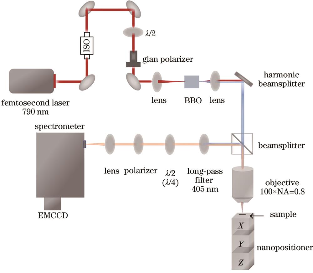
图 2. 单颗粒偏振光谱表征系统原理图
Fig. 2. Schematic of single-particle polarization spectrum characterization system
2.4 偏振光谱检测及分析方法
实验上采用两种偏振检测或分析方法,分别为偏振庞加莱球法和偏振拟合法。对于偏振庞加莱球法,根据6个偏振基矢计算得出的3个归一化斯托克斯参量(S1、S2、S3),可以描述光的任意偏振态。
式中:
偏振拟合法需要在0°~180°范围内,每间隔7.5°旋转半波片快轴记录光强,对应0°~360°范围内偏振角每隔15°探测光强。利用下式拟合随偏振角变化的探测光强,可得参数A、B、
式中:
3 结果分析与讨论
3.1 NaYF4∶Sm3+单颗粒晶体偏振发光测量结果
在395 nm激光的激发下,晶体中Sm3+离子在可见光范围内拥有多个跃迁发光带。实验中,我们测量了544~655 nm范围内光学跃迁的偏振特性。该范围包含3个跃迁发光带,分别为4G5/2-6H5/2(544~570 nm)、4G5/2-6H7/2(570~620 nm)和4G5/2-6H9/2(620~655 nm)[4]。每个跃迁带均由多个晶体场能级跃迁峰组成,由于室温下声子-电子-光子耦合效应[45-47],这些晶体场能级跃迁峰在光谱上很难单独区分。
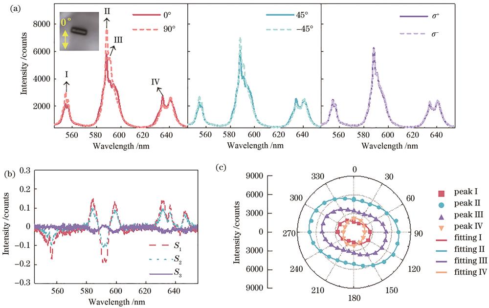
图 3. 六方相NaYF4∶Sm3+单颗粒晶体偏振发光分析。(a)6个偏振庞加莱球基矢下NaYF4∶Sm3+单颗粒光致发光图谱(插图:所测单颗粒面内取向,长约5 μm,0°为光谱仪狭缝方向);(b)所测光谱范围内的S1、S2、S3;(c)4个发光峰强度随偏振角的变化及拟合曲线图谱
Fig. 3. Polarized luminescence analysis of a single hexagonal NaYF4∶Sm3+ microcrystal. (a) Photoluminescence spectra of a single NaYF4∶Sm3+ microcrystal recorded at the six Poincaré sphere basis (inset: measured single-particle in-plane orientation, approximately 5 μm in length, 0° is the slit direction of the spectrometer); (b) S1, S2, and S3 within the measured spectral range; (c) changes in intensity of four emission peaks with polarization angle and fitting curve graph
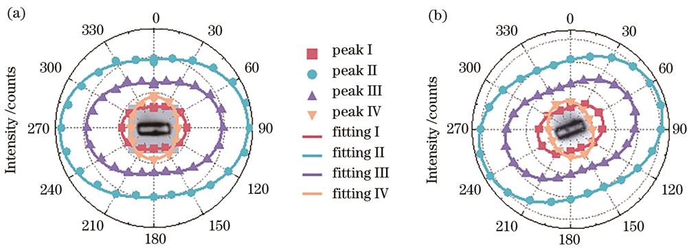
图 4. 两个不同取向的NaYF4∶Sm3+单颗粒晶体的发光峰强度随偏振角的变化及拟合曲线图谱
Fig. 4. Changes in luminescence peak intensity with polarization angle and fitting curve graph of two NaYF4∶Sm3+ single-particle crystals with different orientations
表 1. NaYF4∶Sm3+单颗粒晶体的偏振发光峰拟合参数
Table 1. Fitting parameters of the polarized peaks of single NaYF4∶Sm3+ microcrystals
| ||||||||||||||||||||||||||||||||||||||||||||||||||||||||||||||||||||
3.2 NaYF4∶Sm3+单颗粒晶体偏振发光分析与讨论
由于所选取的4个发光峰均由多个不可区分的晶体场能级跃迁峰交叠而成,为了解释其部分偏振发光特性及线偏角的取向,首先需要了解各晶体场能级跃迁峰的偏振发光特性。在六方相NaYF4∶Sm3+晶体中,Sm3+离子处于Cs微观结晶点群[44],该点群的选择定则允许晶体场能级间产生以电/磁偶极子形式振荡的光学跃迁[41]。由于六方相单颗粒晶体绕结晶c轴具有宏观旋转对称性,发光Sm3+离子的电/磁偶极子也满足旋转对称分布,这意味着同一晶体场能级跃迁峰对应的偶极子与c轴具有相同的极角[28]。在非相干发光情况下,这些跃迁偶极子叠加产生线偏角平行(极角较小)或垂直于c轴(极角较大)的部分线偏跃迁峰。因此,若对任意晶体场能级跃迁峰进行面内线偏测量,探测光强可以表示为
式中:
可以看出,若
3.3 BiPO4∶Sm3+单颗粒晶体偏振发光测量结果
本文利用相同的偏振测试和拟合方法对单斜相BiPO4∶Sm3+单颗粒进行偏振光谱分析。
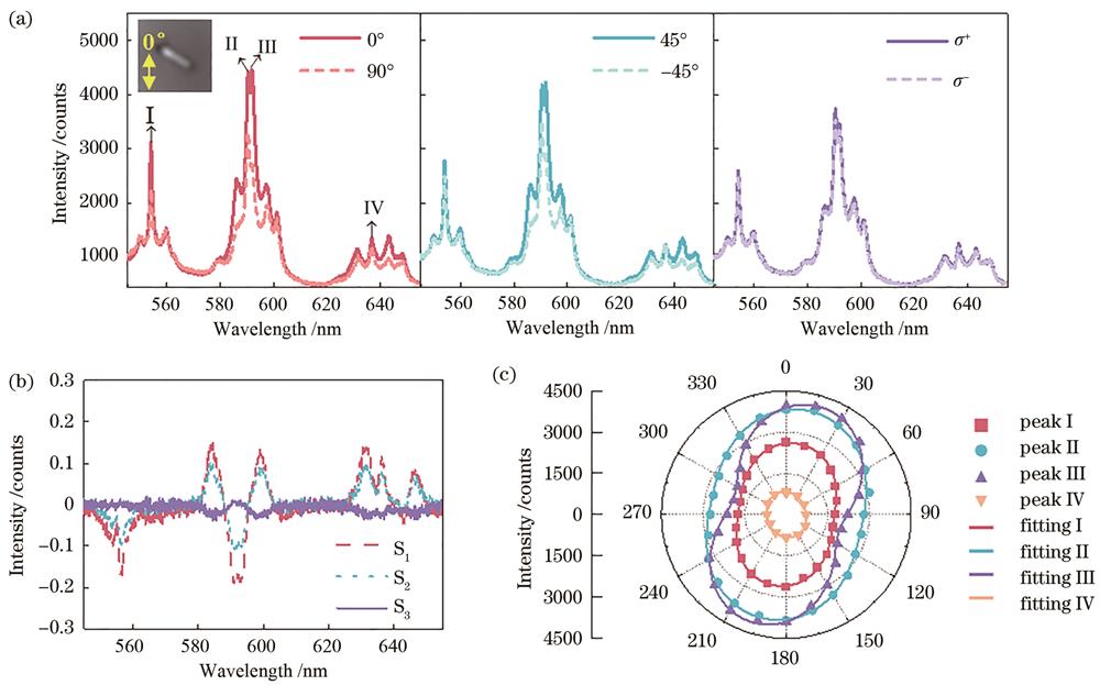
图 5. 单斜相BiPO4∶Sm3+单颗粒晶体偏振发光分析。(a)6个偏振庞加莱球基矢下BiPO4∶Sm3+单颗粒光致发光图谱(插图:所测单颗粒面内取向,长约3 μm);(b)所测光谱范围内的S1、S2、S3;(c)4个发光峰强度随偏振角的变化及拟合曲线图谱
Fig. 5. Polarized luminescence analysis of a single monoclinic BiPO4∶Sm3+ microcrystal: (a) Photoluminescence spectra of a single BiPO4∶Sm3+ microcrystal recorded at the six Poincaré sphere basis (inset: measured single-particle in-plane orientation, approximately 3 μm in length); (b) S1, S2, and S3 within the measured spectral range; (c) changes in intensity of four emission peaks with polarization angle and fitting curve graph
在另外两个不同面内取向的单颗粒晶体上,也得到了相同的偏振测量结论,如
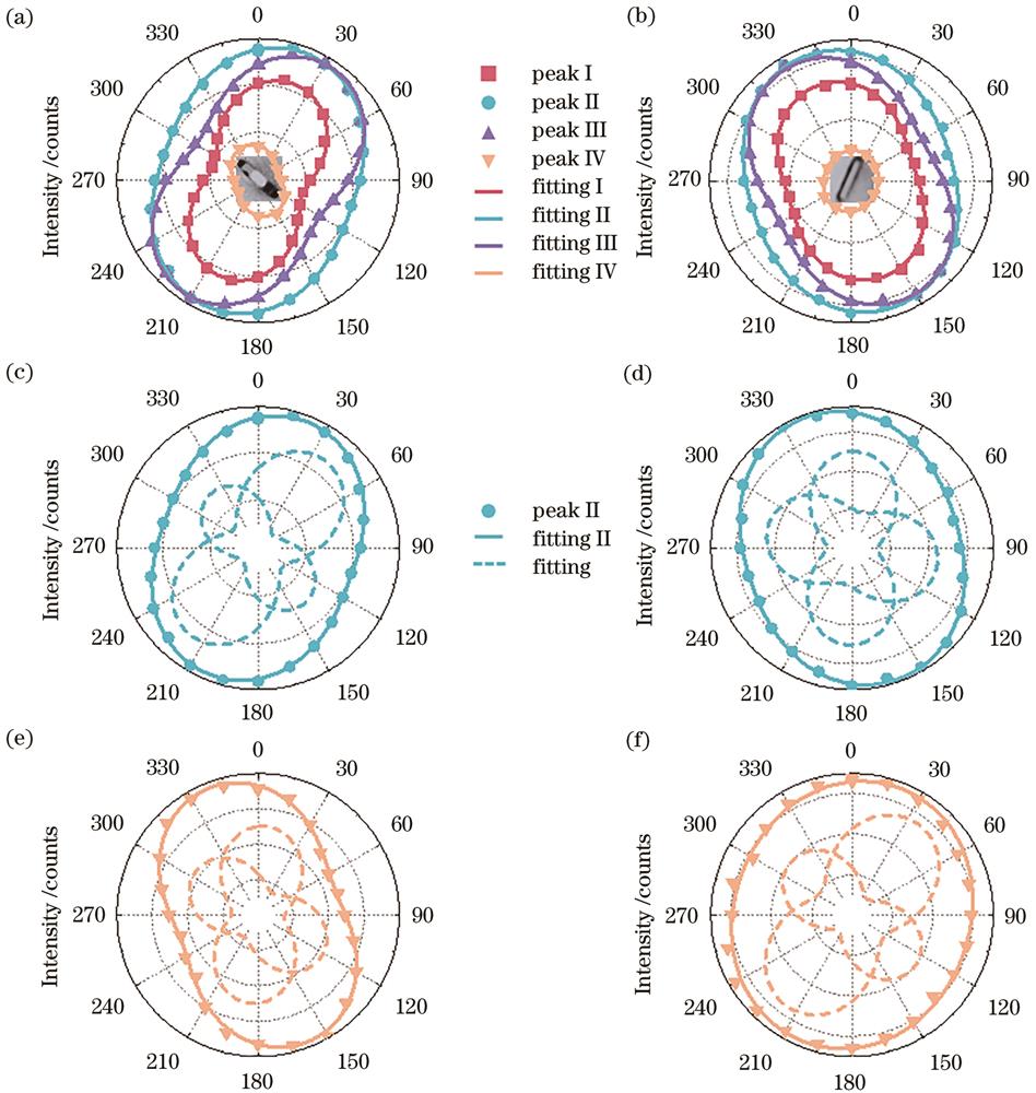
图 6. 两个BiPO4∶Sm3+单颗粒晶体偏振发光拟合分析。(a)(b)单颗粒面内取向和4个发光峰强度随偏振角的变化及拟合曲线图谱;(c)(d)2个不同线偏角的晶体场能级跃迁峰强度叠加拟合发光峰II;(e)(f)2个不同线偏角的晶体场能级跃迁峰强度叠加拟合发光峰IV
Fig. 6. Polarized luminescence analysis of two single BiPO4∶Sm3+ microcrystals. (a) (b) In-plane orientations and changes in intensity of four emission peaks with polarization angle and fitting curve graph; (c) (d) emission peak II fitted by two crystal-field transition peaks with different linear polarization angles; (e) (f) emission peak IV fitted by two crystal-field transition peaks with different linear polarization angles
表 2. BiPO4∶Sm3+单颗粒晶体的偏振发光峰拟合参数
Table 2. Fitting parameters of the polarized peaks of single BiPO4∶Sm3+ microcrystals
| ||||||||||||||||||||||||||||||||||||||||||||||||||||||||||||||||||||
3.4 BiPO4∶Sm3+单颗粒晶体偏振发光分析与讨论
在单斜相BiPO4∶Sm3+晶体中,Sm3+离子处于C1结晶点群[43],该点群的选择定则允许晶体场能级间产生电/磁偶极子振荡光学跃迁,且同一跃迁峰的偶极子振荡方向分别沿着局部点群坐标系的x、y、z轴[41]。由于单斜相单颗粒晶体在宏观上不具备旋转对称性,所有x、y、z取向的偶极子也均不满足绕晶体c轴的旋转对称分布,所以,它们非相干发光形成的部分线偏跃迁峰的线偏角并不沿着或垂直于c轴,而取决于x、y、z偶极子的相对振荡强度。实验上选取的4个发光峰由多个不可区分的晶体场能级跃迁峰交叠而成,为了分析方便,采用2个不同线偏角的跃迁峰拟合实验发光峰,便可以得到很好的拟合效果,如
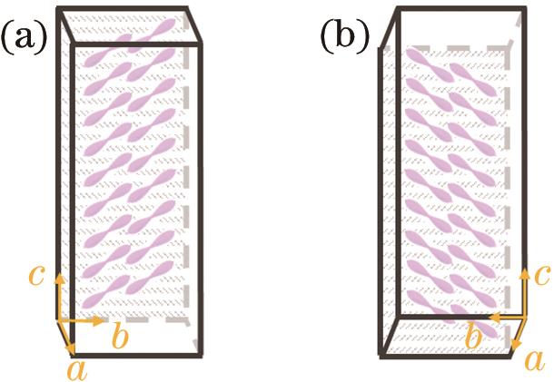
图 7. 单斜相晶体反转导致偶极子沿c轴镜像对称。(a)晶体背面贴附于基板面;(b)晶体正面贴附于基板面
Fig. 7. Reversal of monoclinic crystal results in the dipole mirror symmetry along the c-axis. (a) Reverse side of the crystal attached to the substrate surface; (b) front of the crystal attached to the substrate surface
对比六方相NaYF4∶Sm3+和单斜相BiPO4∶Sm3+单颗粒的偏振发光特性,可以得出结论:无论稀土发光离子处于晶体中何种点群对称性,只要晶体具有三重及以上宏观旋转对称性,稀土单颗粒晶体任意发光峰的线偏角一定精确沿着或垂直于旋转对称轴。
4 结论
本文利用高精度单颗粒偏振光谱表征技术,测量了六方相NaYF4∶Sm3+和单斜相BiPO4∶Sm3+单颗粒晶体发光的偏振特性,发现六方相和单斜相的发光峰均呈现部分偏振特性,且六方相发光峰的线偏角精确沿着或垂直于晶体c轴,单斜相的线偏角与c轴存在一定夹角。基于微观点群理论和晶体的宏观旋转对称性,六方相特殊线偏角起源于光学跃迁偶极子的旋转对称分布,而单斜相不具备旋转对称性导致其发光线偏角随机取向。本研究揭示了稀土单颗粒晶体结构、形貌、取向与其偏振发光间的联系,对于构建具有理想偏振发光特性的稀土单颗粒晶体具有指导意义,有望拓展稀土偏振发光单颗粒在微纳光学传感领域的应用,尤其在传感具有旋转自由度的微观事件方面,例如,纳米物体自组装、微流体局部动力学、细胞内物质转运和生物大分子折叠等。
[1] Zhong T, Kindem J M, Bartholomew J G, et al. Nanophotonic rare-earth quantum memory with optically controlled retrieval[J]. Science, 2017, 357(6358): 1392-1395.
[2] Wang L G, Zhou H P, Hu J N, et al. A Eu3+-Eu2+ ion redox shuttle imparts operational durability to Pb-I perovskite solar cells[J]. Science, 2019, 363(6424): 265-270.
[3] Serrano D, Kuppusamy S K, Heinrich B, et al. Ultra-narrow optical linewidths in rare-earth molecular crystals[J]. Nature, 2022, 603(7900): 241-246.
[4] Ou X Y, Qin X, Huang B L, et al. High-resolution X-ray luminescence extension imaging[J]. Nature, 2021, 590(7846): 410-415.
[5] Liu Y J, Lu Y Q, Yang X S, et al. Amplified stimulated emission in upconversion nanoparticles for super-resolution nanoscopy[J]. Nature, 2017, 543(7644): 229-233.
[6] Lee C, Xu E Z, Liu Y W, et al. Giant nonlinear optical responses from photon-avalanching nanoparticles[J]. Nature, 2021, 589(7841): 230-235.
[7] Kindem J M, Ruskuc A, Bartholomew J G, et al. Control and single-shot readout of an ion embedded in a nanophotonic cavity[J]. Nature, 2020, 580(7802): 201-204.
[8] Han S Y, Deng R R, Gu Q F, et al. Lanthanide-doped inorganic nanoparticles turn molecular triplet excitons bright[J]. Nature, 2020, 587(7835): 594-599.
[9] Fernandez-Bravo A, Yao K Y, Barnard E S, et al. Continuous-wave upconverting nanoparticle microlasers[J]. Nature Nanotechnology, 2018, 13(7): 572-577.
[10] 王浩, 王红宇, 何亮, 等. 新型光功能稀土配合物研究及应用进展[J]. 发光学报, 2022, 43(10): 1509-1523.
[11] Zheng W, Huang P, Tu D T, et al. Lanthanide-doped upconversion nano-bioprobes: electronic structures, optical properties, and biodetection[J]. Chemical Society Reviews, 2015, 44(6): 1379-1415.
[12] Zheng B Z, Fan J Y, Chen B, et al. Rare-earth doping in nanostructured inorganic materials[J]. Chemical Reviews, 2022, 122(6): 5519-5603.
[13] Zeng Z C, Xu Y S, Zhang Z S, et al. Rare-earth-containing perovskite nanomaterials: design, synthesis, properties and applications[J]. Chemical Society Reviews, 2020, 49(4): 1109-1143.
[14] Qin X, Liu X W, Huang W, et al. Lanthanide-activated phosphors based on 4f-5d optical transitions: theoretical and experimental aspects[J]. Chemical Reviews, 2017, 117(5): 4488-4527.
[15] Marin R, Jaque D. Doping lanthanide ions in colloidal semiconductor nanocrystals for brighter photoluminescence[J]. Chemical Reviews, 2021, 121(3): 1425-1462.
[16] Zhou J J, Chizhik A I, Chu S, et al. Single-particle spectroscopy for functional nanomaterials[J]. Nature, 2020, 579(7797): 41-50.
[17] Zhou J J, Chen G X, Wu E, et al. Ultrasensitive polarized up-conversion of Tm3+-Yb3+ doped β-NaYF4 single nanorod[J]. Nano Letters, 2013, 13(5): 2241-2246.
[18] Wei S Q, Shang X Y, Huang P, et al. Polarized upconversion luminescence from a single LiLuF4∶Yb3+/Er3+ microcrystal for orientation tracking[J]. Science China Materials, 2022, 65(1): 220-228.
[19] Rodríguez-Sevilla P, Labrador-Páez L, Wawrzyńczyk D, et al. Determining the 3D orientation of optically trapped upconverting nanorods by in situ single-particle polarized spectroscopy[J]. Nanoscale, 2016, 8(1): 300-308.
[20] Lü Z Y, Dong H, Yang X F, et al. Highly polarized upconversion emissions from lanthanide-doped LiYF4 crystals as spatial orientation indicators[J]. The Journal of Physical Chemistry Letters, 2021, 12(46): 11288-11294.
[21] Kumar A, Kim J, Lahlil K, et al. Optical trapping and orientation-resolved spectroscopy of europium-doped nanorods[J]. Journal of Physics: Photonics, 2020, 2(2): 025007.
[23] Green K K, Wirth J, Lim S F. Nanoplasmonic upconverting nanoparticles as orientation sensors for single particle microscopy[J]. Scientific Reports, 2017, 7: 762.
[24] Chacon R, Leray A, Kim J, et al. Measuring the magnetic dipole transition of single nanorods by spectroscopy and Fourier microscopy[J]. Physical Review Applied, 2020, 14(5): 054010.
[25] Li P, Li F, Zhang X Y, et al. Orthogonally polarized luminescence of single bismuth phosphate microcrystal doped with europium[J]. Advanced Optical Materials, 2020, 8(17): 2000583.
[26] 杨丹丹, 董国平, 邱建荣. 稀土离子掺杂材料的光偏振特性研究进展[J]. 激光与光电子学进展, 2021, 58(15): 1516017.
[27] 邓泽宇, 杨小涵, 张锦文, 等. 稀土上转换发光微纳材料的光物理研究[J]. 中国激光, 2023, 50(1): 0113005.
[28] Li P, Guo Y X, Liu A, et al. Deterministic relation between optical polarization and lattice symmetry revealed in ion-doped single microcrystals[J]. ACS Nano, 2022, 16(6): 9535-9545.
[29] Kim J, Chacón R, Wang Z J, et al. Measuring 3D orientation of nanocrystals via polarized luminescence of rare-earth dopants[J]. Nature Communications, 2021, 12(1): 1943.
[30] Deng K R, Huang X, Liu Y L, et al. Supercrystallographic reconstruction of 3D nanorod assembly with collectively anisotropic upconversion fluorescence[J]. Nano Letters, 2020, 20(10): 7367-7374.
[31] Kim J, Lahlil K, Gacoin T, et al. Measuring the order parameter of vertically aligned nanorod assemblies[J]. Nanoscale, 2021, 13(16): 7630-7637.
[32] Kim J, Michelin S, Hilbers M, et al. Monitoring the orientation of rare-earth-doped nanorods for flow shear tomography[J]. Nature Nanotechnology, 2017, 12(9): 914-919.
[33] Blanc J, Ross D L. Polarized absorption and emission in an octacoordinate chelate of Eu3+[J]. The Journal of Chemical Physics, 1965, 43(4): 1286-1289.
[34] Brecher C. Europium in the ultraphosphate lattice: polarized spectra and structure of EuP5O14[J]. The Journal of Chemical Physics, 1974, 61(6): 2297-2315.
[35] Brecher C, Samelson H, Lempicki A, et al. Polarized spectra and crystal-field parameters of Eu+3 in YVO4[J]. Physical Review, 1967, 155(2): 178-187.
[36] Brecher C, Samelson H, Riley R, et al. Polarized spectra and crystal-field parameters of Eu3+ in YPO4[J]. The Journal of Chemical Physics, 1968, 49(7): 3303-3311.
[37] Sayre E V, Freed S. Absorption spectrum and quantum states of the praseodymium ion. II. anhydrous praseodymium fluoride in films[J]. The Journal of Chemical Physics, 1955, 23(11): 2066-2068.
[38] Sayre E V, Freed S. Spectra and quantum states of the europic ion in crystals. I. absorption spectrum of anhydrous europic chloride[J]. The Journal of Chemical Physics, 1956, 24(6): 1211-1212.
[39] Sayre E V, Freed S. Spectra and quantum states of the europic ion in crystals. II. fluorescence and absorption spectra of single crystals of europic ethylsulfate nonahydrate[J]. The Journal of Chemical Physics, 1956, 24(6): 1213-1219.
[40] Sayre E V, Sancier K M, Freed S. Absorption spectrum and quantum states of the praseodymium ion. I. single crystals of praseodymium chloride[J]. The Journal of Chemical Physics, 1955, 23(11): 2060-2065.
[41] Görller-Walrand C, Binnemans K. Rationalization of crystal-field parametrization[J]. Handbook on the Physics and Chemistry of Rare Earths, 1996, 23: 121-283.
[42] Zhang Y H, Huang L, Liu X G. Unraveling epitaxial habits in the NaLnF4 system for color multiplexing at the single-particle level[J]. Angewandte Chemie, 2016, 128(19): 5812-5816.
[43] Li P, Yuan T L, Li F, et al. Phosphate ion-driven BiPO4∶Eu phase transition[J]. The Journal of Physical Chemistry C, 2019, 123(7): 4424-4432.
[44] Tu D T, Liu Y S, Zhu H M, et al. Breakdown of crystallographic site symmetry in lanthanide-doped NaYF4 crystals[J]. Angewandte Chemie International Edition, 2013, 52(4): 1128-1133.
[45] Mishra S K, Gupta M K, Ningthoujam R S, et al. Presence of water at elevated temperatures, structural transition, and thermal expansion behavior in LaPO4∶Eu[J]. Physical Review Materials, 2018, 2(12): 126003.
[46] Liang F, He C, Lu D Z, et al. Multiphonon-assisted lasing beyond the fluorescence spectrum[J]. Nature Physics, 2022, 18(11): 1312-1316.
[47] Li Z H, Hudry D, Heid R, et al. Phonon density of states in lanthanide-based nanocrystals[J]. Physical Review B, 2020, 102(16): 165409.
Article Outline
岳新, 叶洳言, 郭雅欣, 李朋, 李峰. Sm3+掺杂NaYF4和BiPO4单颗粒微米晶体发光偏振特性研究[J]. 激光与光电子学进展, 2023, 60(11): 1106028. Xin Yue, Ruyan Ye, Yaxin Guo, Peng Li, Feng Li. Polarized Luminescence of Sm3+-Doped Single NaYF4 and BiPO4 Microcrystals[J]. Laser & Optoelectronics Progress, 2023, 60(11): 1106028.
