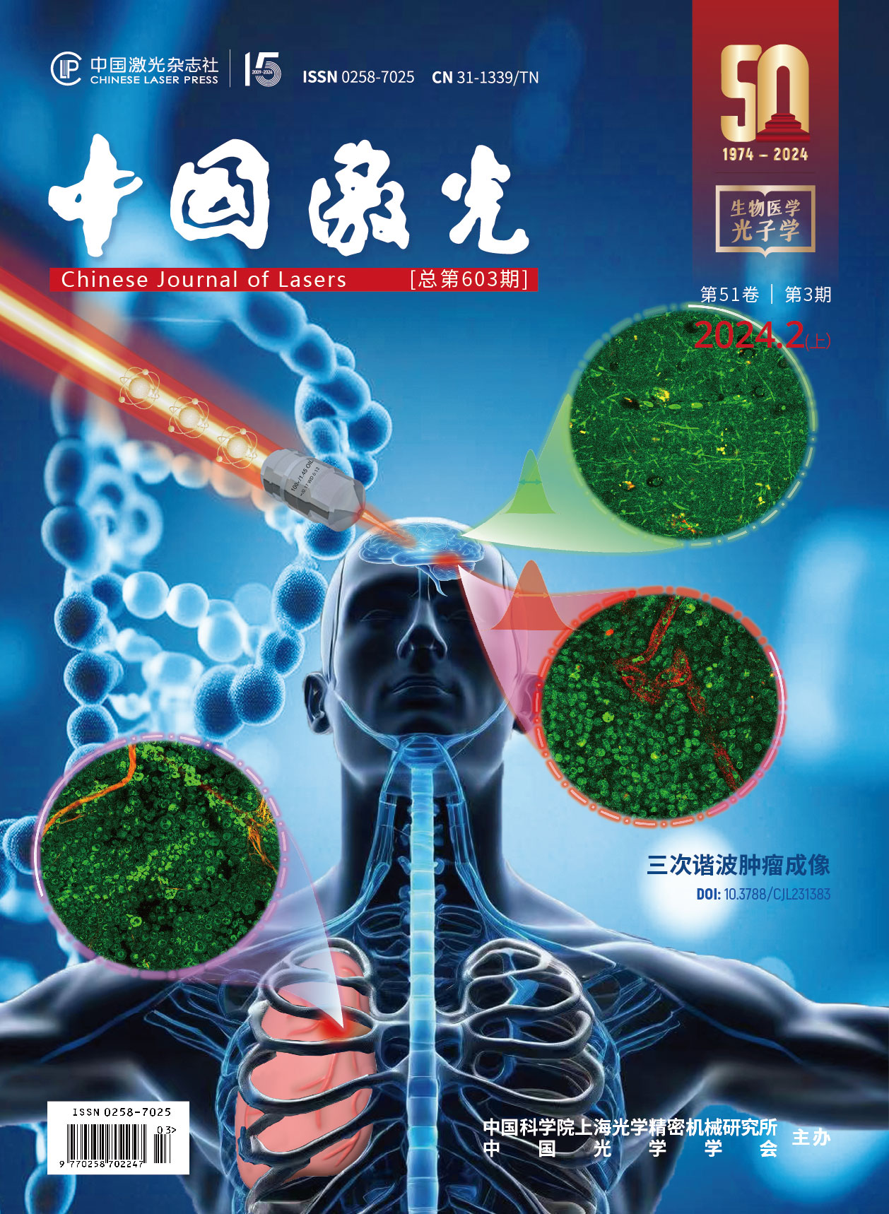
三次谐波显微成像在肿瘤诊断中的应用进展封面文章
")
Cancer remains a major life-threatening disease worldwide, as reported by the World Health Organization (WHO). Surgery is the primary therapy for most solid tumors, with the ideal outcome relying on a balance between complete tumor removal and maximal preservation of surrounding normal tissue. Current clinical imaging modalities such as magnetic resonance imaging (MRI), computed tomography (CT), and positron emission tomography (PET) lack the resolution to accurately delineate tumor boundaries. The gold standard in clinics for detecting tumor boundaries and infiltration is the histopathological analysis of surgical specimens via hematoxylin and eosin (H&E) staining. However, the H&E staining workflow requires time-consuming tissue processing, including formalin fixation, paraffin embedding, and manual staining, often taking more than a day before diagnostic results are available to surgeons. Consequently, there is an urgent demand for new real-time microscopic imaging techniques that can be used intraoperatively to provide instant feedback during tumor surgery.
Recent years have seen promising developments in label-free nonlinear imaging techniques for real-time tissue pathology in the operating room. These techniques include multiphoton fluorescence microscopy, optical coherence tomography (OCT), Raman microscopy, and harmonic microscopy, which can visualize tumor margins without exogenous labels. Among these, third harmonic generation (THG) combined with second harmonic generation (SHG) offers a unique, label-free subcellular-resolution assessment of fresh and unprocessed tissues. THG signals arise from nonlinear three-photon optical responses at cell-cell and cell-matrix interfaces (Fig. 1), effectively detecting proliferative cells and vasculatures, key hallmarks of tumor pathology. THG microscopy stands out by providing sub-cellular resolution, rich cellular and molecular information, and images of H&E quality. Additionally, using a single beam, complementary information from SHG, two-photon excited fluorescence (2PEF), and three-photon excited fluorescence (3PEF) can be simultaneously collected, visualizing extensive architectural and molecular details. These advantages position THG imaging as a highly promising technique for intraoperative determination of tumor margins.
In this review, we explore the fundamental principles of the THG nonlinear process and discuss its latest applications in intraoperative tumor imaging. We highlight recent engineering innovations enabling miniaturized, portable THG imaging systems suitable for operating room deployment. We also review pioneering efforts in developing THG-capable endoscope probes using flexible fiber-optics, potentially integrating with standard surgical equipment. Embedding THG microscopy seamlessly into clinical workflows can provide surgeons with real-time, in-situ histopathology, enhancing surgical outcomes without disrupting the surgical rhythm. This review aims to accelerate the translation and adoption of label-free nonlinear optical imaging, particularly THG microscopy, as a valuable intraoperative guidance tool.
Recent studies have demonstrated the potential of integrated THG, SHG, and multiphoton fluorescence microscopy for ex-vivo characterization of freshly resected human brain tumors (Fig. 2), ovarian tumors, breast cancer specimens, lung tumors (Fig. 3), and other tumor types. These studies reveal pathological hallmarks such as increased cellularity, nuclear pleomorphism, and vascular proliferation. The in-situ extraction of tumor pathological features underscores THG imaging's potential to improve surgical outcomes. Efforts are underway to transition THG microscopy from benchtop to clinically viable tools. Most THG microscopes are currently confined to research labs, with large volumes, complex opto-mechanical components, and limited consideration for patient safety or imaging stability. To facilitate widespread intraoperative use, miniaturized and portable THG imaging platforms are necessary. Researchers in the Netherlands and the USA have independently developed compact, multimodal THG microscopes, and these devices have been tested in clinical settings, such as operation rooms or pathological laboratories, for pilot clinical validation (Fig. 4). These devices enable on-site assessment of surgical specimens and provide rapid diagnostic feedback for tumor classification and margin determination, assisting surgeons in decision-making. However, existing miniaturized THG microscopes are limited to ex-vivo imaging. To enable real-time, in-situ guidance without tissue removal, endoscopic techniques are essential for THG imaging. The nonlinear imaging field is witnessing increasing efforts to design THG-capable endoscopes, drawing from innovations in 2PEF/3PEF, SHG, OCT, and Raman microscopy (Fig. 5). THG endoscopy is still in its early stages, presenting numerous opportunities for scientific research, technology translation, and clinical studies.
THG imaging shows promise for real-time intraoperative assessment of various cancer types. Significant progress has been made in developing compact, portable THG imaging systems for intraoperative use. Currently, only two groups have begun clinical testing with their portable THG microscopes. More systematic clinical testing is needed to further mature this technology for routine operation room use. Additionally, technical translations from other imaging modalities are required to advance THG endoscopy solutions. Despite the vast potential of THG microscopy for real-time, non-destructive assessment of fresh tissue, more efforts from both the scientific and industrial sectors are imperative to promote the translation of THG microscopes from laboratories to clinical settings.
薄启宇, 吴宇辰, 邱斯奇, 张志清. 三次谐波显微成像在肿瘤诊断中的应用进展[J]. 中国激光, 2024, 51(3): 0307101. Qiyu Bo, Yuchen Wu, Siqi Qiu, Zhiqing Zhang. Current Progress of Third Harmonic Generation Microscopy in Tumor Diagnosis[J]. Chinese Journal of Lasers, 2024, 51(3): 0307101.


 PDF全文
PDF全文

