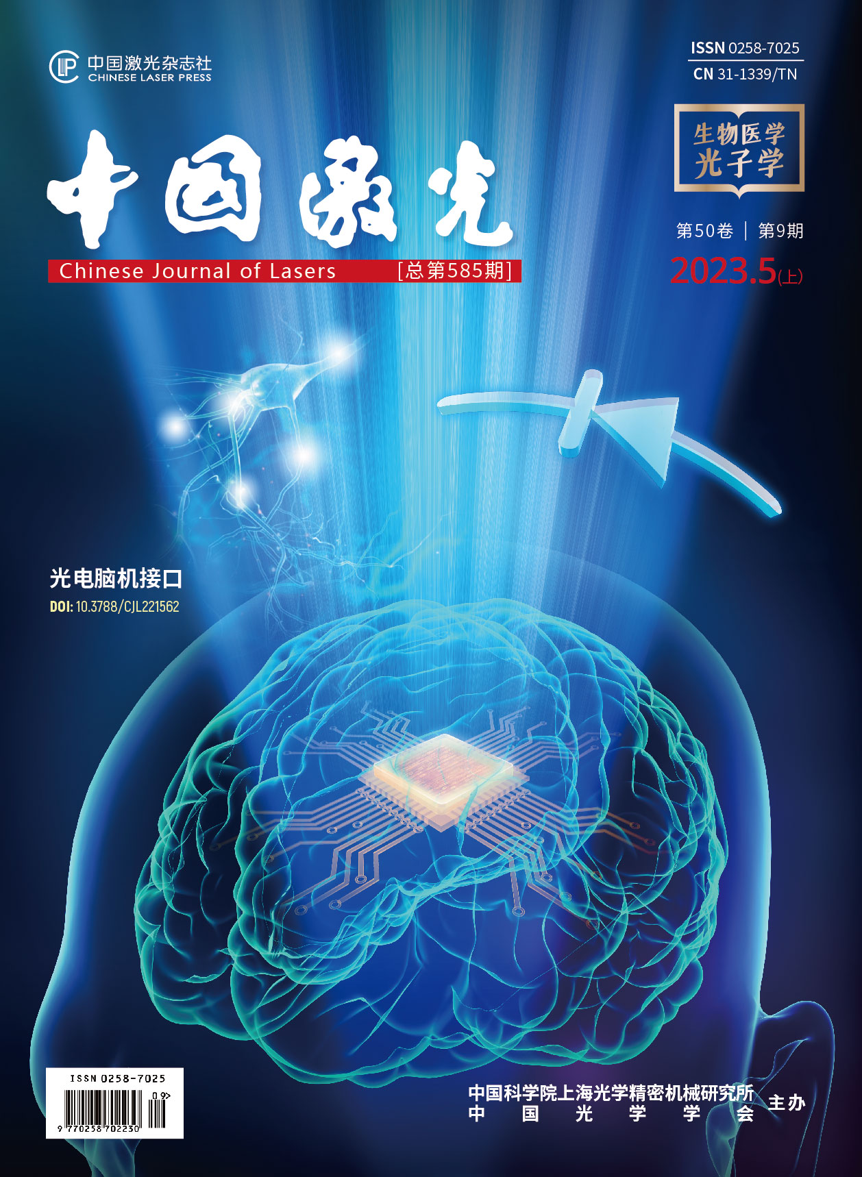
利用深度学习扩展双光子成像视场  下载: 502次
李迟件, 姚靖, 高玉峰, 赖溥祥, 何悦之, 齐苏敏, 郑炜. 利用深度学习扩展双光子成像视场[J]. 中国激光, 2023, 50(9): 0907107.
Chijian Li, Jing Yao, Yufeng Gao, Puxiang Lai, Yuezhi He, Sumin Qi, Wei Zheng. Extending Field‑of‑View of Two‑Photon Microscopy Using Deep Learning[J]. Chinese Journal of Lasers, 2023, 50(9): 0907107.
[1] Zipfel W R, Williams R M, Webb W W. Nonlinear magic: multiphoton microscopy in the biosciences[J]. Nature Biotechnology, 2003, 21(11): 1369-1377.
[2] König K. Multiphoton microscopy in life sciences[J]. Journal of Microscopy, 2000, 200(2): 83-104.
[3] Helmchen F, Denk W. Deep tissue two-photon microscopy[J]. Nature Methods, 2005, 2(12): 932-940.
[4] Bumstead J R, Park J J, Rosen I A, et al. Designing a large field-of-view two-photon microscope using optical invariant analysis[J]. Neurophotonics, 2018, 5(2): 025001.
[5] Ji N, Freeman J, Smith S L. Technologies for imaging neural activity in large volumes[J]. Nature Neuroscience, 2016, 19(9): 1154-1164.
[6] Yang W J, Yuste R. In vivo imaging of neural activity[J]. Nature Methods, 2017, 14(4): 349-359.
[7] Tsai P S, Mateo C, Field J J, et al. Ultra-large field-of-view two-photon microscopy[J]. Optics Express, 2015, 23(11): 13833-13847.
[8] Yu C H, Stirman J N, Yu Y Y, et al. Diesel2p mesoscope with dual independent scan engines for flexible capture of dynamics in distributed neural circuitry[J]. Nature Communications, 2021, 12(1): 1-8.
[9] Clough M, Chen I A, Park S W, et al. Flexible simultaneous mesoscale two-photon imaging of neural activity at high speeds[J]. Nature Communications, 2021, 12(1): 1-7.
[10] Demas J, Manley J, Tejera F, et al. High-speed, cortex-wide volumetric recording of neuroactivity at cellular resolution using light beads microscopy[J]. Nature Methods, 2021, 18(9): 1103-1111.
[11] Sofroniew N J, Flickinger D, King J, et al. A large field of view two-photon mesoscope with subcellular resolution for in vivo imaging[J]. eLife, 2016, 5: e14472.
[12] Stirman J N, Smith I T, Kudenov M W, et al. Wide field-of-view, multi-region two-photon imaging of neuronal activity in the mammalian brain[J]. Nature Biotechnology, 2016, 34(8): 857-62.
[13] Yao J, Gao Y F, Yin Y X, et al. Exploiting the potential of commercial objectives to extend the field of view of two-photon microscopy by adaptive optics[J]. Optics Letters, 2022, 47(4): 989-992.
[14] 刘立新, 张美玲, 吴兆青, 等. 自适应光学在荧光显微镜中的应用[J]. 激光与光电子学进展, 2020, 57(12): 120001.
[15] Booth M J, Neil M A A, Juskaitis R, et al. Adaptive aberration correction in a confocal microscope[J]. Proceedings of the National Academy of Sciences of the United States of America, 2002, 99(9): 5788-5792.
[16] Park J H, Kong L J, Zhou Y F, et al. Large-field-of-view imaging by multi-pupil adaptive optics[J]. Nature Methods, 2017, 14(6): 581-583.
[17] Ji N. Adaptive optical fluorescence microscopy[J]. Nature Methods, 2017, 14(4): 374-380.
[18] Booth M J. Adaptive optical microscopy: the ongoing quest for a perfect image[J]. Light: Science & Applications, 2014, 3(4): e165.
[19] Zhou H, Cai R Y, Quan T W, et al. 3D high resolution generative deep-learning network for fluorescence microscopy imaging[J]. Optics Letters, 2020, 45(7): 1695-1698.
[20] Zhang H, Fang C Y, Xie X L, et al. High-throughput, high-resolution deep learning microscopy based on registration-free generative adversarial network[J]. Biomedical Optics Express, 2019, 10(3): 1044-1063.
[21] Weigert M, Schmidt U, Boothe T, et al. Content-aware image restoration: pushing the limits of fluorescence microscopy[J]. Nature Methods, 2018, 15(12): 1090-1097.
[22] Cheng S F, Zhou Y Y, Chen J B, et al. High-resolution photoacoustic microscopy with deep penetration through learning[J]. Photoacoustics, 2022, 25: 100314.
[23] Zhao H X, Ke Z W, Chen N B, et al. A new deep learning method for image deblurring in optical microscopic systems[J]. Journal of Biophotonics, 2020, 13(3): e201960147.
[24] Belthangady C, Royer L A. Applications, promises, and pitfalls of deep learning for fluorescence image reconstruction[J]. Nature Methods, 2019, 16(12): 1215-1225.
[25] 李浩宇, 曲丽颖, 华子杰, 等. 基于深度学习的荧光显微成像技术及应用[J]. 激光与光电子学进展, 2021, 58(18): 1811007.
[26] Liu J H, Huang X S, Chen L Y, et al. Deep learning–enhanced fluorescence microscopy via degeneration decoupling[J]. Optics Express, 2020, 28(10): 14859-14873.
[27] 熊子涵, 宋良峰, 刘欣, 等. 基于深度学习的荧光显微性能提升(特邀)[J]. 红外与激光工程, 2022, 51(11): 89-106.
Xiong Z H, Song L F, Liu X, et al. Performance enhancement of fluorescence microscopy by using deep learning(invited)[J]. Infrared and Laser Engineering, 2022, 51(11): 89-106.
[28] Shen B L, Liu S W, Li Y P, et al. Deep learning autofluorescence-harmonic microscopy[J]. Light: Science & Applications, 2022, 11(1): 1-14.
[29] Wang Z Q, Zhu L X, Zhang H, et al. Real-time volumetric reconstruction of biological dynamics with light-field microscopy and deep learning[J]. Nature Methods, 2021, 18(5): 551-556.
[32] Zheng Y, Chen J J, Wu C X, et al. Adaptive optics for structured illumination microscopy based on deep learning[J]. Cytometry, 2021, 99(6): 622-631.
[34] Hu L J, Hu S W, Gong W, et al. Image enhancement for fluorescence microscopy based on deep learning with prior knowledge of aberration[J]. Optics Letters, 2021, 46(9): 2055-2058.
[35] RonnebergerO, FischerP, BroxT. U-Net: convolutional networks for biomedical image segmentation[M]∥Navab N, Hornegger J, Wells W M, et al. Medical image computing and computer-assisted intervention-MICCAI 2015. Lecture notes in computer science. Cham: Springer, 2015: 234-241.
[36] HeK M, ZhangX Y, RenS Q, et al. Deep residual learning for image recognition[C]∥2016 IEEE Conference on Computer Vision and Pattern Recognition (CVPR), June 27-30, 2016, Las Vegas, NV, USA. New York: IEEE Press, 2016: 770-778.
[39] nBRAnet[EB/OL]. [2022-10-08]. https://gitee.com/li-chijian/nBRAnet.
[40] KimJ, LeeJ K, LeeK M. Accurate image super-resolution using very deep convolutional networks[C]∥2016 IEEE Conference on Computer Vision and Pattern Recognition (CVPR), June 27-30, 2016, Las Vegas, NV, USA. New York: IEEE Press, 2016: 1646-1654.
李迟件, 姚靖, 高玉峰, 赖溥祥, 何悦之, 齐苏敏, 郑炜. 利用深度学习扩展双光子成像视场[J]. 中国激光, 2023, 50(9): 0907107. Chijian Li, Jing Yao, Yufeng Gao, Puxiang Lai, Yuezhi He, Sumin Qi, Wei Zheng. Extending Field‑of‑View of Two‑Photon Microscopy Using Deep Learning[J]. Chinese Journal of Lasers, 2023, 50(9): 0907107.


