Author Affiliations
Abstract
1 Integrative Oncology Department – Imaging Unit, BC Cancer Research Institute, Vancouver, BC, Canada
2 Department of Dermatology and Skin Science, University of British Columbia and Vancouver Coastal Health Research Institute, Vancouver, BC, Canada
Multi-photon microscopy (MPM) and coherent anti-Stokes Raman scattering (CARS) are two advanced nonlinear optical imaging techniques, which provide complementary information and have great potential in combination for noninvasive in vivo biomedical applications. This paper provides a detailed discussion of the basics, development and applications of these technologies for in vivo skin research, covering the following topics: The principle and advantage of MPM and CARS, instrumentation development for in vivo applications, MPM and CARS of normal skin, application of MPM and CARS in skin cancer and disease diagnosis; application of MPM in skin disease intervention, i.e., imaging guided two-photon photothermolysis.Multi-photon microscopy (MPM) and coherent anti-Stokes Raman scattering (CARS) are two advanced nonlinear optical imaging techniques, which provide complementary information and have great potential in combination for noninvasive in vivo biomedical applications. This paper provides a detailed discussion of the basics, development and applications of these technologies for in vivo skin research, covering the following topics: The principle and advantage of MPM and CARS, instrumentation development for in vivo applications, MPM and CARS of normal skin, application of MPM and CARS in skin cancer and disease diagnosis; application of MPM in skin disease intervention, i.e., imaging guided two-photon photothermolysis.
Nonlinear microscopy multiphoton microscopy coherent anti-Stokes Raman scattering microscopy skin skin cancer multiphoton therapy Journal of Innovative Optical Health Sciences
2023, 16(1): 2230018

Author Affiliations
Abstract
1 Departamento de Física, Universidade Federal de Pernambuco, Recife-PE 50670-901, Brazil
2 Instituto de Física, Universidade Federal de Goiás, Goiânia-GO 74001-970, Brazil
The confocal microscopy technique was applied for nonlinear optical characterization of single β-barium-borate (β-BBO) nanocrystals. The experimental setup allows measurements of the laser polarization-selective second-harmonic (SH) generation, and the results can be used to determine the nanocrystals’ c-axis orientation, as well as to obtain information about their second-order susceptibility χ(2). The dependence of the SH signal on the laser polarization allowed the discrimination of individual particles from aggregates. The data were fitted using a model that takes into account the BBO properties and the experimental setup characteristics considering (i) the electrostatic approximation, (ii) the effects of the microscope objective used to focus the light on the sample in an epi-geometry configuration, and (iii) the symmetry of χ(2) for the β-BBO nanocrystals. A signal at the third-harmonic frequency was also detected, but it was too weak to be studied in detail.
190.3970 Microparticle nonlinear optics 180.4315 Nonlinear microscopy Chinese Optics Letters
2018, 16(4): 041902
Author Affiliations
Abstract
1 College of Physics and Energy, Shenzhen University, P. R. China, 518060
2 Key Laboratory of Optoelectronic Devices and Systems of Ministry of Education and Guangdong Province, College of Optoelectronic Engineering, Shenzhen University , Shenzhen, P. R. China, 518060
Optical microscopy of biological tissues at the 1700 nm window has enabled deeper penetration, due to the combined advantage of relatively small water absorption and tissue scattering at this wavelength. Compared with excitation at other wavelengths, such as the commonly used 800 nm window for two-photon microscopy, water absorption at the 1700 nm window is more than one order of magnitude higher. As a result, more temperature rise can be expected and can be potentially detrimental to biological tissues. Here, we present theoretical estimation of temper-ature rise at the focus of objective lens at the 1700 nm window, purely due to water absorption. Our calculated result shows that under realistic experimental conditions, temperature rise due to water absorption is still below 1 K and may not cause tissue damage during imaging.
Nonlinear microscopy multiphoton processes temperature Journal of Innovative Optical Health Sciences
2017, 10(2): 1650048
Author Affiliations
Abstract
1 Britton Chance Center for Biomedical Photonics, School of Engineering Sciences, Wuhan National Laboratory for Optoelectronics–Huazhong University of Science and Technology, Wuhan 430074, China
2 MoE Key Laboratory for Biomedical Photonics, Department of Biomedical Engineering, Huazhong University of Science and Technology, Wuhan 430074, China
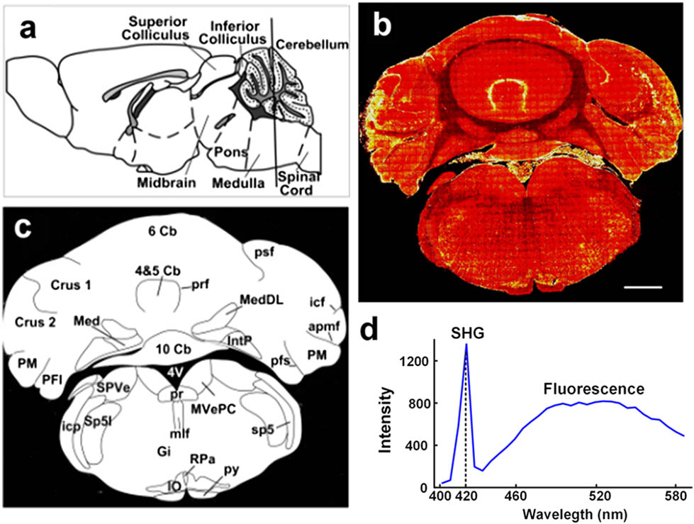
To visualize the structure and organization of the brain is a fundamental requirement in the research of neuroscience. Here, combining with two-photon excitation fluorescence microscopy and transgenetic mouse GAD67, we demonstrate a custom-built second harmonic generation (SHG) microscope to discriminate brain layers and sub regions in the cerebellum and brain stem slices with cellular resolution. In particular, the cell densities of neurons in different brain layers are extracted due to the cell soma appearing as dark shadow on an SHG image. Further, the axon initial segments of the Purkinje cell are easily recognized without labeling, which would be useful for guiding micropipettes for electrophysiology.
170.3880 Medical and biological imaging 180.4315 Nonlinear microscopy Chinese Optics Letters
2017, 15(9): 090003
1 华中科技大学武汉光电国家实验室(筹)Britton Chance生物医学光子学研究中心, 湖北 武汉 430074
2 华中科技大学生物医学工程系生物医学光子学教育部重点实验室, 湖北 武汉 430074
双光子荧光显微成像是一种非线性光学显微技术,具有高空间分辨率、高信噪比和固有的三维层析分辨能力等优点。传统的双光子荧光显微成像通常使用波长可调谐的100 fs超短脉冲激光器作为激光光源。目前,人们对双光子荧光显微成像方法进行了深入研究,改进光源及探测方法是常用的手段。介绍和总结了多色双光子荧光显微成像技术的近期研究进展及其在生物医学中的应用。首先介绍了传统飞秒激光器及光学参量振荡器在多色成像中的应用,然后对光纤超连续谱在多色显微成像中的应用进行了分析,最后简要说明了增强自相位调制效应产生连续光谱以及选择性激发实现多色成像的工作。多色双光子成像技术不仅可以同时获取含有多种荧光团的待测样品的高对比度双光子荧光图像,而且具有系统结构简单、操作简便等优点,这使得其在生物医学和材料科学等领域具有广阔的应用前景,并且为生物医学诊断与研究提供了一种有效的工具和平台。
显微 非线性显微成像 双光子荧光显微成像 多色成像 超连续谱 激光与光电子学进展
2017, 54(6): 060002
Author Affiliations
Abstract
1 Leibniz Institute of Photonic Technology (IPHT-Jena) Albert Einstein Straβe 9, 07745 Jena, Germany
2 Institute of Physical Chemistry and Abbe Center of Photonics Friedrich Schiller University Jena, Helmholtzweg 4, 07743 Jena, Germany
3 National Institute of Optics, National Research Council (INO-CNR) Largo E. Fermi 6 – 50125, Florence, Italy
4 European Laboratory for Non-Linear Spectroscopy (LENS) University of Florence Via Nello Carrara 1-50019, Sesto Fiorentino (Firenze), Italy
5 Clinic for Internal Medicine, Jena University Hospital Friedrich-Schiller-University, Erlanger Allee 101, 07747 Jena, Germany
6 Institute of Pathology, Department of Neuropathology Jena University Hospital – Friedrich-Schiller-University Erlanger Allee 101, 07740 Jena, Germany
7 Catholic Clinic — Koblenz, Internal Medicine & Cardiology Rudolf Virchow Str. 9, 56073 Koblenz, Germany
Cardiovascular diseases in general and atherothrombosis as the most common of its individual disease entities is the leading cause of death in the developed countries. Therefore, visualization and characterization of inner arterial plaque composition is of vital diagnostic interest, especially for the early recognition of vulnerable plaques. Established clinical techniques provide valuable morphological information but cannot deliver information about the chemical composition of individual plaques. Therefore, spectroscopic imaging techniques have recently drawn considerable attention. Based on the spectroscopic properties of the individual plaque components, as for instance different types of lipids, the composition of atherosclerotic plaques can be analyzed qualitatively as well as quantitatively. Here, we compare the feasibility of multimodal nonlinear imaging combining two-photon fluorescence (TPF), coherent anti-Stokes Raman scattering (CARS) and second-harmonic generation (SHG) microscopy to contrast composition and morphology of lipid deposits against the surrounding matrix of connective tissue with diffraction limited spatial resolution. In this contribution, the spatial distribution of major constituents of the arterial wall and atherosclerotic plaques like elastin, collagen, triglycerides and cholesterol can be simultaneously visualized by a combination of nonlinear imaging methods, providing a powerful label-free complement to standard histopathological methods with great potential for in vivo application.
Nonlinear microscopy Raman spectroscopy atherosclerosis Journal of Innovative Optical Health Sciences
2014, 7(5): 1450027
Author Affiliations
Abstract
In the past two decades, two-photon microscopy (TPM) transforms biomedical research, allowing nondestructive high-resolution fluorescent molecular imaging and label-free imaging in vivo and in real time. Here we review the recent advances of TPM technology including novel laser sources, new image acquisition paradigms, and microendoscopic imaging systems. Then, we survey the capabilities of TPM imaging of biological relevant molecules such as nicotinamide adenine dinucleotide (NADH), flavin adenine dinucleotide (FAD), and reactive oxygen species (ROS). Biomedical applications of TPM in neuroscience and cancer detection are demonstrated.
170.3880 Medical and biological imaging 170.0170 Medical optics and biotechnology 180.4315 Nonlinear microscopy 180.0180 Microscopy 170.2150 Endoscopic imaging 170.0170 Medical optics and biotechnology 190.4180 Multiphoton processes 190.0190 Nonlinear optics 170.6510 Spectroscopy, tissue diagnostics 170.0170 Medical optics and biotechnology Chinese Optics Letters
2013, 11(1): 011703
福建师范大学物理与光电信息科技学院, 福建 福州 350108
光学显微镜的发展历史是一段不断提高显微镜的分辨率和对比度的历史。双光子显微镜是近30年来非线性显微镜的研究发展的代表。它在分辨率上与共聚焦显微镜相当, 但在成像的层析穿透深度上有显著提高, 并且大大减少了光毒性与光漂白。由于生物细胞组织中富有各种自家荧光源, 因此双光子显微镜被广泛应用于皮肤组织甚至癌组织以及细胞的成像。基于共聚焦扫描显微镜的双光子显微镜可以很容易的与二次谐波显微镜组合, 对皮肤组织中的重要成分胶原纤维进行成像。双光子显微镜还可以结合其他非线性光学现象对组织以及细胞进行成像, 显示其强大的生命力。将来随着携带方便且廉价的双光子显微镜的出现, 双光子显微镜有望在临床医学上发挥其有效的作用。
双光子显微镜 非线性显微镜 自家荧光源 two photon microscopy nonlinear microscopy autofluorescenct fluorophore
Author Affiliations
Abstract
1 Institute of Biophotonics, National Yang-Ming University, 155, Linong St., Sec. 2, Taipei 112, China
2 Institute of Microbiology and Immunology, National Yang-Ming University, 155, Linong St., Sec. 2, Taipei 112, China
3 Department of Photonics, National Sun Yat-Sen University, 70 Lien Hai Road, Kaohsiung 804, China
Fluorescence lifetime imaging microscopy (FLIM) has gained popularity as a sensitive technique to monitor the functional/conformational states of reduced nicotinamide adenine dinucleotide (NADH), one of the main compounds of oxidative phosphorylation. In this letter, we apply the technique to characterize the metabolic changes in mouse embryonic fibroblast 3T3 cells upon bacterial infection. A gradual shortening of the decaying time constants in both the short and the long lifetime components of NADH’s autofluorescence is detected. The ratio of the short and the long lifetime components’ relative contributions, however, shows a rapid increase, indicating the rise of cellular metabolic activity over the course of infection.
细菌感染 NADH 荧光寿命成像 000.1430 Biology and medicine 000.4920 Other life sciences 110.1080 Active or adoptive optics 180.2520 Fluorescence microscopy 180.4315 Nonlinear microscopy 300.2530 Fluorescence, laser-induced Chinese Optics Letters
2010, 8(10): 931
Author Affiliations
Abstract
Key Laboratory of OptoElectronic Science and Technology for Medicine, Ministry of Education, Fujian Provincial Key Laboratory of Photonic Technology, School of Physics and OptoElectronics Technology, Fujian Normal University, Fuzhou 350007, China
Two-photon excitation fluorescence (TPEF) and second-harmonic generation (SHG) are detected through multiphoton microscopy (MPM). The major signals have the potential to monitor the process of tissue changes. TPEF and SHG are used to monitor the skin photo-thermal response to irradiation with intense pulsed light sources (\lambda is in the range of 560?1200 nm) and trace the process of skin remodeling in vivo at different time intervals. TPEF intensity is nearly unchanged at different time intervals after irradiation, whereas SHG intensity changes considerably. The results reveal the photo-thermal effect of nonablative light sources and the process of collagen remodeling at the sub-micron level.
光热响应 修复 多光子显微镜 170.1870 Dermatology 170.3880 Medical and biological imaging 180.4315 Nonlinear microscopy Chinese Optics Letters
2010, 8(8): 784


