1 广州医科大学生物医学工程学院医学影像创新实验室,广东 广州 511436
2 广州医科大学附属第一医院呼吸疾病国家重点实验室,广东 广州 510120
巨噬细胞作为炎症阶段的主要吞噬细胞,其高表达是急性呼吸道炎症发展过程的临床特征之一。目前还没有一种成像方法能够以深组织穿透性和高分辨率的方式呈现巨噬细胞在急性炎症中的表达。以吲哚菁绿纳米颗粒(Nano-ICG)作为一种高效的光声成像(PAI)增强造影剂,评估了急性呼吸道炎症中巨噬细胞的表达量。激光共聚焦显微镜下的成像效果证实,Nano-ICG能够快速地被巨噬细胞吞噬。利用Nano-ICG增强光声成像效果后,气管内的PAI结果显示了巨噬细胞在炎症后气管壁上的分布区域。Nano-ICG增强的光声成像能够无创、定量地评估急性呼吸道炎症的发展程度,有望为呼吸疾病相关基础研究和临床诊疗提供新的影像技术支持。
医用光学 光声成像 急性呼吸道炎症 吲哚菁绿纳米颗粒 巨噬细胞
1 Prokhorov General Physics Institute of the Russian Academy of Sciences, Moscow 119991, Russia
2 National Research Nuclear University MEPhI, Moscow 115409, Russia
3 Institute of Gene Biology, Russian Academy of Sciences, Moscow 119334, Russia
4 Burdenko Neurosurgical Institute, Moscow 125047, Russia
5 Serbsky National Medical Research Centre of Psychiatry and Narcology under the RF Ministry of Public Health, Moscow 119034, Russia
Frontiers of Optoelectronics
2020, 13(4): 371
1 湖南师范大学蛋白质化学与发育生物学教育部重点实验室, 长沙 410081
2 湖南省南华生物医药研究所, 长沙 410015
3 中南大学湘雅三医院, 长沙 410013
炎症性疾病的发生是当今临床医学攻克的重点。M1型巨噬细胞分泌炎症因子产生炎症, 而M2型巨噬细胞分泌抑炎因子抑制炎症的发生。M1型巨噬细胞向M2型极化, 则是从炎症状态转变成抑制炎症发生的状态, 因此研究巨噬细胞向缓解炎症的M2型极化将有利于炎症性疾病的治疗。本研究利用骨髓间充质干细胞(BMSC)培养液处理已被脂多糖(LPS)诱导呈M1型的Raw264.7巨噬细胞, 探究骨髓间充质干细胞培养液(BMSC-CM)对巨噬细胞向M2型极化的影响及其分子机制。提取来源于3周龄C57BL/6鼠的骨髓间充质干细胞; 再收集BMSC-CM处理M1型的Raw264.7巨噬细胞; 半定量PCR检测M1型标记基因[肿瘤坏死因子α(TNF-α)和诱导型一氧化氮合酶(INOS)]和M2型标记基因[精氨酸酶1(ARG-1)和转化生长因子β1(TGF-β1)]mRNA表达以及白介素10(IL-10)mRNA表达水平; Western蛋白质印迹法检测信号传导及转录激活蛋白3(STAT3)和磷酸化STAT3(p-STAT3)的表达。本研究发现,经过BMSC-CM培养后的M1型的Raw264.7巨噬细胞, 其M2型相关指标ARG-1和TGF-β1 mRNA水平明显上升, 并且IL-10 mRNA水平和p-STAT3蛋白水平也明显上升。这些结果说明,骨髓间充质干细胞培养液通过IL-10/STAT3信号通路促进STAT3磷酸化, 诱导巨噬细胞Raw264.7细胞向M2型极化。
骨髓间充质干细胞 巨噬细胞极化 M1/M2表型 bone marrow-derived mesenchymal stem cell macrophage polarization macrophages M1/M2 phenotyp STAT3 STAT3 IL-10 IL-10
Author Affiliations
Abstract
1 State Key Laboratory of Molecular Vaccinology and Molecular Diagnostics & Center for Molecular Imaging and Translational Medicine, School of Public Health, Xiamen University, Xiamen 361102, China
2 State Key Laboratory of Precision Electronic Manufacturing Technology and Equipment, School of Electromechanical Engineering, Guangdong University of Technology, Guangzhou 510006, China
3 Jiangxi Key Laboratory of Optic-Electronic and Communication, Jiangxi Science and Technology Normal University, Nanchang 330038, China
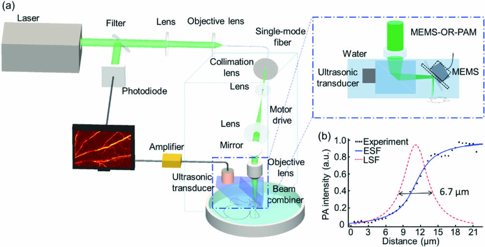
Photoacoustic imaging has been developed to image the immune study at the macro scale. Macrophages play diverse roles in the acute response to infection and tissue repair. However, macrophages activities in acute inflammation at the microscopic level still remain challenging. In this work, we proposed optical-resolution photoacoustic microscopy to promptly monitor the labeled macrophages activities in normal and inflammatory groups. The result showed that many labeled macrophages emerged around the vessels firstly, then exuded into tissues, and finally disappeared in the inflammatory group injected with labeled macrophages. In summary, our method allows us to exactly image and track the immune cells of inflammatory diseases.
photoacoustic microscopy macrophages activities vessel parameter Chinese Optics Letters
2020, 18(12): 121701
Author Affiliations
Abstract
1 Interdisciplinary Center of Critical Technologies in Medicine, Saratov State University, 83 Astrakhanskaya Str. Saratov 410012, Russia
2 Institute of Biochemistry and Physiology of Plants and Microorganisms, Russian Academy of Sciences, 13 Entuziastov Ave. Saratov 410049, Russia
3 Saratov State Medical University, 112 Bolshaya Kazachia Str., Saratov 410012, Russia
4 Department of Optics and Biophotonics, Saratov State University, 83 Astrakhanskaya Str. Saratov 410012, Russia
5 Yuri Gagarin State Technical University of Saratov, 77 Politechnicheskaya Str., Saratov 410054, Russia
6 Laboratory of Laser Diagnostics of Technical and Living Systems Institute of Precision, Mechanics and Control of RAS, 24 Rabochaya Str., Saratov 410028, Russia
7 Laboratory of Biophotonics, Tomsk State University, 36 Lenin's Ave. Tomsk 634050, Russia
Malignant gliomas are highly invasive tumors that use the cerebral vessels for invasion due to high vascular fragility of the blood–brain barrier (BBB). On one hand, glioma is characterized by the BBB disruption, on the other hand, drug brain delivery via the BBB is a big challenge in glioma therapy. The limited information about vascular changes associated with glioma growth is a reason of slow progress in prevention of glioma development. Here, we present in vivo and ex vivo study of the BBB disruption and glioma cells (GCs) migration in rats using fluorescence and confocal microscopy. We uncovered a local breach in the BBB in the main tumor mass but not within the border of normal and malignant cells, where the BBB was impermeable for high weight molecules. The migration of GCs were observed via the cerebral vessels with the intact BBB that was associated with macrophages infiltration. The mechanisms underlying glioma progression remain unknown but there is an evidence that the sympathetic nervous system (SNS) via activation of vascular beta2-adrenoreceptors (B2-ADRs) can play an important role in tumor metastasis. Our results clearly show an increase in the expression of vascular B2-ADRs and production of the beta-arrestin-1 - co-factor of B2-ADRs signaling pathway in rats with glioma. Pharmacological blockade of B2-ADRs reduces the BBB disruption, macrophages infiltration, GCs migration and increases survival rate. These data suggest that the blockade of B2-ADRs may be a novel adjuvant therapeutic strategy to reduce glioma progression and prevent metastasis.
Glioma macrophages blood–brain barrier beta-2-adrenoreceptors beta-arrestin-1 Journal of Innovative Optical Health Sciences
2018, 11(4): 1850025
Author Affiliations
Abstract
1 Department of Chemistry, Fudan University, Shanghai, P. R. China
2 Institutes of Biomedical Sciences, Fudan University, Shanghai, P. R. China
3 Department of Urology, Xinhua Hospital Shanghai Jiao Tong University 1665, Kongjiang Road, Shanghai 200092, P. R. China
4 Med-X Research Institute, Shanghai Jiao Tong University 1954, Huashan Road, Shanghai 200240, P. R. China
5 School of Biomedical Engineering Shanghai Jiao Tong University, Shanghai, P. R. China
Metastasis is a very complicated multi-step process and accounts for the low survival rate of the cancerous patients. To metastasize, the malignant cells must detach from the primary tumor and migrate to secondary sites in the body through either blood or lymph circulation. Macrophages appear to be directly involved in tumor progression and metastasis. However, the role of macrophages in affecting cancer metastasis has not been fully elucidated. Here, we have utilized an emerging technique, namely in vivo flow cytometry (IVFC) to study the depletion kinetics of circulating prostate cancer cells in mice and determine how depletion of macrophages by the liposome-encapsulated clodronate affects the depletion kinetics. Our results show different depletion kinetics of PC-3 cells between the macrophage-deficient group and the control group. The number of circulating tumor cells (CTCs) in the macrophage-deficient group decreases in a slower manner compared to the control mice group. The differences in depletion kinetics indicate that the absence of macrophages facilitates the stay of prostate cancer cells in circulation. In addition, our imaging data suggest that macrophages might be able to arrest, phagocytose and digest PC-3 cells. Therefore, phagocytosis may mainly contribute to the depletion kinetic differences. The developed methods elaborated here would be useful to study the relationship between macrophages and tumor metastasis in small animal cancer models.
Prostate cancer macrophages liposome-encapsulated clodronate in vivo flow cytometer circulating tumor cells Journal of Innovative Optical Health Sciences
2012, 5(4): 1250027
华南师范大学激光生命科学研究所,广东 广州 510631
用He-Ne激光辐照巨噬细胞,实时观测活体单细胞内钙浓度([Ca2+]i)分布和细胞免疫活性随激光功率和照射时间的变化.实验结果表明,随激光照射时间的增加,[Ca2+]i出现上升达到最大值后又下降,逐渐回复初始状态,且呈现中心最强,径向衰减的环形梯度分布.不同激光剂量对巨噬细胞内[Ca2+]i及其免疫活性有着不同的影响,同一波长激光,当剂量不同时,可以表现为完全相反的效应.同时激光功率也是影响巨噬细胞内[Ca2+]i及其免疫活性的一个重要因素,同一波长、同一剂量情况下,如果激光功率不同,巨噬细胞内[Ca2+]i及其免疫活性的变化也有着显著的差异.当照射功率为0.16 mW时,胞内[Ca2+]i峰值是照射功率为0.40 mW时的近6倍.最大值时对应细胞免疫活性最高.对其机理作了初步探讨.
免疫病理学 激光共焦扫描显微镜 He-Ne激光 巨噬细胞 钙浓度([Ca2+]i) 免疫活性


