Author Affiliations
Abstract
1 State Key Laboratory of Modern Optical Instrumentations, Centre for Optical and Electromagnetic Research, College of Optical, Science and Engineering, International Research Center for Advanced Photonics, Zhejiang University, Hangzhou 310058, P. R. China
2 Dr. Li Dak Sum & Yip Yio Chin Center for Stem Cell and Regenerative Medicine, Zhejiang University, Hangzhou 310058, P. R. China
3 College of Biomedical Engineering and Instrument Science, Interdisciplinary Institute of Neuroscience and Technology (ZIINT), Zhejiang University, Hangzhou 310027, P. R. China
Fluorescence imaging in the second near-infrared window (NIR-II, 900–1880nm) with less scattering background in biological tissues has been combined with the confocal microscopic system for achieving deep in vivo imaging with high spatial resolution. However, the traditional NIR-II fluorescence confocal microscope with separate excitation focus and detection pinhole makes it possess low confocal efficiency, as well as difficultly to adjust. Two types of upgraded NIR-II fluorescence confocal microscopes, sharing the same pinhole by excitation and emission focus, leading to higher confocal efficiency, are built in this work. One type is fiber-pinhole-based confocal microscope applicable to CW laser excitation. It is constructed for fluorescence intensity imaging with large depth, high stabilization and low cost, which could replace multiphoton fluorescence microscopy in some applications (e.g., cerebrovascular and hepatocellular imaging). The other type is air-pinhole-based confocal microscope applicable to femtosecond (fs) laser excitation. It can be employed not only for NIR-II fluorescence intensity imaging, but also for multi-channel fluorescence lifetime imaging to recognize different structures with similar fluorescence spectrum. Moreover, it can be facilely combined with multiphoton fluorescence microscopy. A single fs pulsed laser is utilized to achieve up-conversion (visible multiphoton fluorescence) and down-conversion (NIR-II one-photon fluorescence) excitation simultaneously, extending imaging spectral channels, and thus facilitates multi-structure and multi-functional observation.
Self-confocal fiber-pinhole air-pinhole multi-channel fluorescence lifetime imaging multi-color imaging Journal of Innovative Optical Health Sciences
2024, 17(1): 2350025
1 滨州学院飞行学院,山东 滨州 256600
2 北京空间机电研究所,北京 100094
3 大连工业大学信息科学与工程学院,辽宁 大连 116034
系统地探究了典型多光谱彩色成像系统的最优光谱通道数的确定问题。在前期多目标滤色片优化选取方法的基础上,将仅限于相同通道滤色器优化的概念拓展至不同通道数最优滤色器的优化,从而达到最优通道数确定的目的。基于Munsell光谱反射率数据集构建光谱反射率成像目标,通过真实CCD成像传感器的光谱灵敏度、D65光源的光谱功率分布以及高斯滤色器模型和最大线性独立滤色器选择算法,在10个噪声水平下实现了3~31个光谱通道,即29个虚拟多光谱相机对成像目标的光谱反射率重建的仿真计算。结果表明,通道数小于8时,5通道滤色器表现最优;和A光源相比,D65光源下的5通道最优滤色器的最大带宽达到80 nm,性能有显著的提升。
色度学 成像系统 计算方法 光谱通道数 多光谱彩色成像
1 中国科学技术大学生物医学工程学院(苏州),江苏 苏州 215163
2 中国科学院苏州生物医学工程技术研究所,江苏 苏州 215163
单像素成像技术以其较强的弱光探测能力和较宽的工作波段在生物医学成像领域有着广阔的应用前景。通过将单像素成像技术与内窥成像技术相结合,提出两种腹腔镜全彩单像素内窥成像技术方案:一种是RGB三色光源方案将颜色和空间信息分别按时序分配给照明和检测双方,另一种是白光光源方案通过三个探测器同时采集颜色和空间的信息。搭建了两种腹腔镜全彩单像素内窥成像系统,设计了模块化的单像素相机,以多色彩条和肠道模型为成像目标,通过实验从峰值信噪比、结构相似度和成像速度等方面,系统量化分析了两种全彩单像素内窥成像系统的技术指标。实验结果表明,两种方案峰值信噪比和结构相似度接近,而白光光源方案成像速度更快,可以适配各种腹腔镜进行内窥成像,为推动单像素成像在内窥成像领域的应用提供了理论指导。
单像素成像 内窥成像 单像素相机 全彩成像 single-pixel imaging endoscopic imaging single-pixel camera full-color imaging 红外与激光工程
2023, 52(10): 20230077
Author Affiliations
Abstract
Shandong Provincial Engineering and Technical Center of Light Manipulations & Shandong Provincial Key Laboratory of Optics and Photonic Device, School of Physics and Electronics, Shandong Normal University, Jinan 250014, China
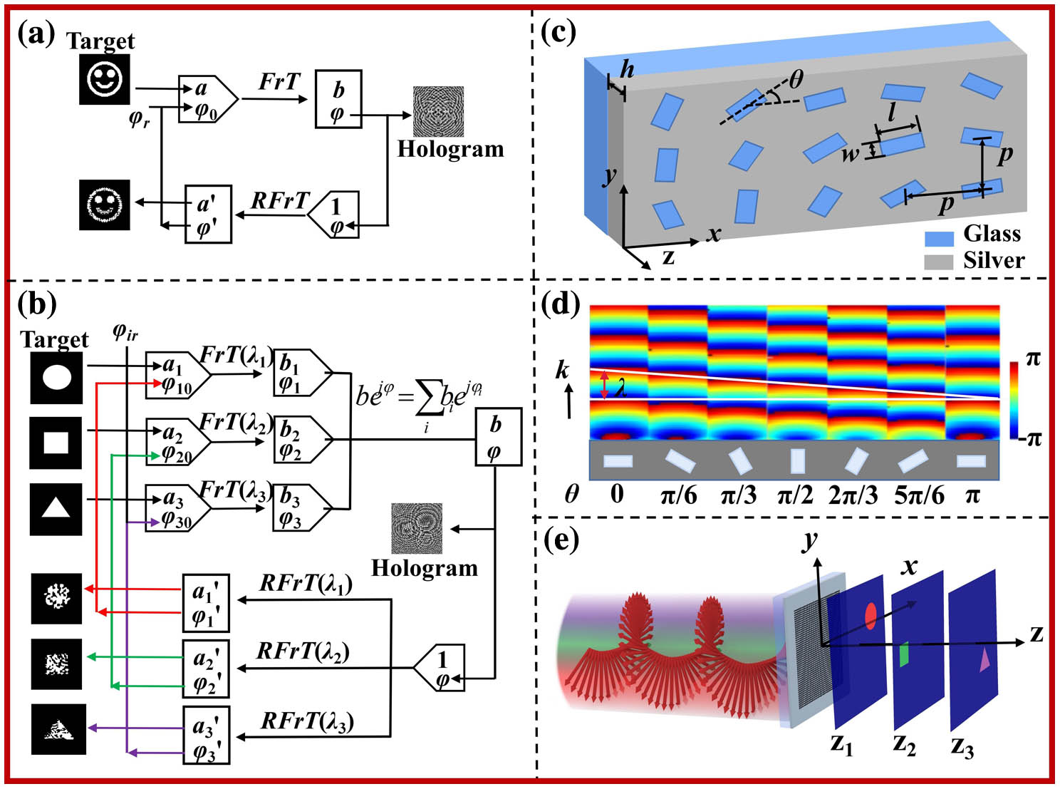
In light of the powerful light manipulation ability of holographic metasurfaces, optical imaging with wavelength multiplexing and polarization multiplexing is performed in this paper. The metasurface is composed of identical rectangular nanoholes etched in silver film. Three imaging effects, including the in-plane color imaging, three-dimensional wavelength-encrypted imaging, and polarization-multiplexing wavelength-encrypted imaging, are realized. The designed metasurface has compact structure, and the obtained image has lower noise. The simulation and experiment results give the verification. Multiple images, including spatial multiplexing, wavelength multiplexing, and polarization multiplexing, exhibit immense potentialities of metasurfaces, and this work is helpful for expanding the applications of metasurfaces.
metasurface holography optical encryption color imaging Chinese Optics Letters
2023, 21(10): 100501
1 海南省生物医学工程重点实验室, 海南大学 生物医学工程学院, 海南 海口 570100
2 海南大学 计算机科学与技术学院, 海南 海口 570100
数字病理凭借其便捷的存储、管理、浏览、传输等特点,为远程病理会诊及联合会诊带来了新契机。然而,显微镜的视场有限,在保证分辨率的前提下,无法兼顾全景成像。全景数字病理的提出弥补了这一缺陷,其在保证分辨率的同时可兼顾全景成像。但单张切片仅能实现单靶点检测,而疾病诊断需同时观测多个靶点的表达情况。近年来,多靶点全景数字病理技术发展迅速,因其在药物研发、临床科研以及基础科研等领域有巨大的应用潜力而广受关注。该系统凭借视场大、颜色多、通量高的特点,可在短时间内原位检测整张组织切片上的多种生物标记物的表达情况,借以识别组织上每个细胞表型、丰度、状态及其相互关系。本文首先梳理了数字病理、全景数字病理以及多靶点全景数字病理的发展过程,并简要介绍发展过程中技术的更新迭代,以及发展多靶点全景数字病理的重要性。然后,分别从生物样本准备、多色光学成像以及图像处理3个部分重点介绍多靶点全景数字病理。接下来,阐述了多靶点全景数字病理在肿瘤微环境与肿瘤分子分型等生物医学领域的应用情况。最后,对多靶点全景数字病理的技术优势、目前面临的挑战及其未来的发展趋势进行了总结。
多靶点全景数字病理 生物标记物 多色成像 图像处理 multi-target panoramic digital pathology biomarkers multi-color imaging image processing 
Author Affiliations
Abstract
1 Nanophotonics Research Center, Shenzhen Key Laboratory of Micro-Scale Optical Information Technology & Institute of Microscale Optoelectronics, Shenzhen University, Shenzhen 518060, China
2 Department of Physics, Harbin Institute of Technology, Harbin 150001, China
We introduce a simple one-dimensional (1D) structure in the design of 1D color splitters (1D-CSs) with RGB unit cells for color imaging and propose a single-to-double-layer design in 1D-CSs. Based on inverse design metasurfaces, we demonstrate numerically a single-layer 1D-CS with a full-color efficiency of 46.2% and a double-layer 1D-CS with a full-color efficiency of 48.2%; both of them are significantly higher than that of traditional color filters. Moreover, we demonstrate a 1D-CS that has application value by evaluating the double-layer 1D-CS’s performances in terms of incident angle sensitivity, polarization angle sensitivity, and assembly tolerance.
color splitter color imaging inverse design metasurfaces Chinese Optics Letters
2022, 20(7): 073601

Author Affiliations
Abstract
1 State Key Laboratory of Optical Technologies on Nano-Fabrication and Micro-Engineering, Institute of Optics and Electronics, Chinese Academyof Sciences, Chengdu 610209, China
2 School of Optoelectronics, University of Chinese Academy of Sciences, Beijing 100049, China
Dispersion control is crucial in optical systems, and chromatic aberration is an important factor affecting imaging quality in imaging systems. Due to the inherent property of materials, dispersion engineering is complex and needs to trade off other aberration in traditional ways. Although metasurface offers an effective method to overcome these limits and results in well-engineered dispersion, off-axis dispersion control is still a challenging topic. In this paper, we design a single-layer metalens which is capable of focusing at three wavelengths (473 nm, 532 nm, and 632 nm) with different incident angles (0°, -17° and 17°) into the same point. We also demonstrate that this metalens can provide an alternative for the bulky color synthetic prism in a 3-chips digital micromirror device (DMD) laser projection system. Through this approach, various off-axis dispersion controlling optical devices could be realized.
off-axis dispersion control metalens color imaging Opto-Electronic Advances
2020, 3(4): 04190005


